. 2013 Sep;25(9):3584-601.
doi: 10.1105/tpc.113.114017. Epub 2013 Sep 30.

The C2H2 transcription factor regulator of symbiosome differentiation represses transcription of the secretory pathway gene VAMP721a and promotes symbiosome development in Medicago truncatula

Affiliations

The C2H2 transcription factor regulator of symbiosome differentiation represses transcription of the secretory pathway gene VAMP721a and promotes symbiosome development in Medicago truncatula

Senjuti Sinharoy et al. Plant Cell. 2013 Sep.

Abstract

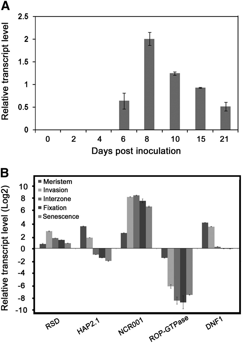
Transcription factors (TFs) are thought to regulate many aspects of nodule and symbiosis development in legumes, although few TFs have been characterized functionally. Here, we describe regulator of symbiosome differentiation (RSD) of Medicago truncatula, a member of the Cysteine-2/Histidine-2 (C2H2) family of plant TFs that is required for normal symbiosome differentiation during nodule development. RSD is expressed in a nodule-specific manner, with maximal transcript levels in the bacterial invasion zone. A tobacco (Nicotiana tabacum) retrotransposon (Tnt1) insertion rsd mutant produced nodules that were unable to fix nitrogen and that contained incompletely differentiated symbiosomes and bacteroids. RSD protein was localized to the nucleus, consistent with a role of the protein in transcriptional regulation. RSD acted as a transcriptional repressor in a heterologous yeast assay. Transcriptome analysis of an rsd mutant identified 11 genes as potential targets of RSD repression. RSD interacted physically with the promoter of one of these genes, VAMP721a, which encodes vesicle-associated membrane protein 721a. Thus, RSD may influence symbiosome development in part by repressing transcription of VAMP721a and modifying vesicle trafficking in nodule cells. This establishes RSD as a TF implicated directly in symbiosome and bacteroid differentiation and a transcriptional regulator of secretory pathway genes in plants.


Figures

Figure 1.
Figure 1.
Protein Sequence, Conserved Domains, and Phylogenetic Analysis of RSD. (A) Alignment of Mt-RSD amino acid sequence with that of putative orthologs in L. japonicus and soybean. Conserved amino acids are highlighted. The C2H2 DNA binding domain and EAR domain are marked. (B) Unrooted phylogenetic unweighted pair group method with arithmetic mean tree of Mt-RSD homologs/orthologs in M. sativa (Ms), Arabidopsis (At), Populus trichocarpa (Pt), G. max (Gm), L. japonicus (Lj), and Oryza sativa (Os). Sequences were identified via BLASTP searches of the National Center for Biotechnology Information database of nonredundant sequences and aligned at the protein level using multiple sequence comparison by log expectation (see Supplemental Data Set 1 online). At-ZAP6, which has a C2H2 DNA binding domain but lacks the EAR domain, was used as an outgroup. Numbers represent the percentage of 1000 bootstrap replications to assess robustness of nodes. The asterisk indicates the nodulation-specific subgroup of proteins. The bar represents estimated amino acid change per sequence position.
Figure 2.
Figure 2.
Temporal and Spatial Expression Profiles of Mt-RSD. (A) Expression profile of RSD during nodule development in M. truncatula genotype R108. qRT-PCR was used to measure RSD transcript levels in the infection-susceptible zone of roots (0 to 4 DAI) and in developing nodules (6 to 21 DAI) (B) Relative transcript levels of RSD in different zones of nodules of genotype A17 at 28 DAI. HAP2.1, NCR001, ROP-GTPase, and DNF1 relative transcript levels are included for comparison. Mean and sd of three biological replicates are presented in each case.
Figure 3.
Figure 3.
Symbiotic Phenotypes of rsd Mutants and Complementation by p35S-GFP-MtRSD. (A) Schematic representation of the Mt-RSD gene with Tnt1 insertion sites (arrowheads) in two independent lines. The black region represents the protein coding region, and UTRs are shown in gray. Bar = 100 bp. (B) Wild-type R108 (left), rsd-1 (NF11265; middle), and rsd-2 (NF3492; right) at 21 DAI with S. meliloti strain 2011. Close-up views of root nodules of the wild type (left), rsd-1 (middle), and rsd-2 (right) are shown below. (C) Acetylene reduction activity (ARA) of whole nodulated roots of R108, rsd-1, and rsd-2 at 15 DAI. Data are the mean of 41 plants for R108, 32 plants for rsd-1, and 12 plants for rsd-2. Vertical bars represent se. n.d., not detected. (D) to (G) Complementation of the rsd-1 (NF11265) mutant with a p35S-GFP-MtRSD construct via Agrobacterium rhizogenes–mediated hairy root transformation. Stereomicroscope images under light ([D] and [F]) and fluorescent illumination ([E] and [G]). Bars = 1 mm. (D) and (E) Spheroidal nodules on control roots transformed with empty vector at 28 DAI. (F) and (G) Cigar-shaped nodules on roots transformed with p35S-GFP-MtRSD at 28 DAI. The inset in (F) shows a typical, pink complemented nodule.
Figure 4.
Figure 4.
Bacterial Colonization Phenotypes of Nodules from Wild-Type and rsd-1 Mutant Plants. Wild-type (R108) nodules ([A] to [C]) and rsd-1 mutant nodules ([D] to [F]) at 6 DAI ([A] and [D]), 8 DAI ([B] and [E]), and 21 DAI ([C] and [F]). Bars = 100 μm in (A), (B), (D), and (E) and 200 μm in (C) and (F). lacZ-expressing S. meliloti strain 2011 cells are blue from X-Gal staining.
Figure 5.
Figure 5.
Differentiation of Bacteroids Is Impaired in rsd-1 Mutant Nodules. (A) to (H) Phenotype of R108 ([A] to [D]) and rsd-1 ([E] to [H]) nodules at 12 DAI with S. meliloti strain 1022 containing the mCherry plasmid. Nodules were stained with FM1-43 (green) to visualize membranes ([A], [C], [E], and [G]). The asterisk in (E) marks autofluorescence. Bars = 100 μm in (A) and (E), 10 μm in (B), (C), (F), and (G), and 1 μm in (D) and (H). (B) and (F) Light micrographs of semithin sections from 12-DAI nodules stained with toluidine blue-O. (B), (C), (F), and (G) Cells of the invasion zone. (D) and (H) Transmission electron micrographs of infected cells showing bacteroids in zone II. (I) to (L) Distribution of ploidy levels of plant and bacteroid cells in nodules at 18 DAI with S. meliloti 1021. (I) and (J) DNA content of plant cells from wild-type R108 and rsd-1 nodules, respectively. DNA content of individual nuclei was estimated by relative DAPI fluorescence. 2C, 4C, 8C, 16C, 32C, and 64C DNA content are indicated. (K) and (L) DNA content of DAPI-stained bacteroids measured by flow cytometry.
Figure 6.
Figure 6.
Expression of RSD in Various Fix Nodules. Relative transcript levels of Mt-RSD in different types of Fix nodules measured at 10 DAI. Asterisks indicate a significant difference in RSD expression with respect to the Fix+ control: *P ≤ 0.05, **P ≤ 0.01, and ***P ≤ 0.001. Mean and sd of three biological replicates are presented in each case. (A) Various Fix plant mutants. (B) Various Fix bacterial mutants in wild-type plants.
Figure 7.
Figure 7.
Mt-RSD Acts as a Transcriptional Repression. (A) Effector and reporter constructs for testing Mt-RSD transcriptional activation and repression activity in yeast. Gal4-DBD, Gal4 DNA binding domain; VP16, activation domain of the VP16 protein; GAL4-DB motif, Gal4-DBD binding site. β-Galactosidase assays showing activation of lacZ expression by each construct. Average and standard deviations are calculated from three independent experiments. (B) Expression profile of Mt-RSD and Mt-VAMP721a. qRT-PCR was used to measure Mt-RSD and Mt-VAMP721a transcript levels at 0, 6, and 8 DAI in M. truncatula R108 and rsd-1 nodules. (C) ChIP followed by quantitative PCR showed a significant enrichment in Medtr4g022570 (Mt-VAMP721a) promoter DNA detected with primer set 2 (pSET2) from RSD-transformed hairy roots compared with controls transformed with the empty vector. The presumptive binding motif is indicated. (D) EMSA of the 288-bp promoter region (fragment −1870 to −1581, with respect ATG) as the probe labeled with biotin in the presence (lanes 2 to 4) or absence (lane 1) of purified His-tagged Mt-RSD. Unlabeled promoter fragment (−1870 to −1581) in 25-fold (lane 3) or 50-fold (lane 4) molar excess relative to the biotin-labeled sequence was used as specific competitor.
Figure 8.
Figure 8.
Hypothetical Mode of Action of Mt-RSD during Nodule Development. IT delivery and endocytosis of rhizobia (green) and biogenesis of symbiosomes in a single plant cell are depicted. Mt-VAMP721d and Mt-VAMP721e are required to target and fuse specialized vesicles, presumably containing cell wall–degrading enzymes, to the IT membrane, which leads to dissolution of the IT cell wall and bacterial endocytosis (Ivanov et al., 2012). These VAMPs may continue to deliver membrane and cargo to the symbiosomes as they proliferate. The endoplasmic reticulum and Golgi are involved in the biogenesis of these and other types of secretory vesicles. Mt-RSD blocks the production of Mt-VAMP721a (via transcriptional repression), eliminating an alternative secretory pathway that may compete with the pathway(s) required for symbiosome biogenesis or alternatively may act in plant defense. Mt-RSD is crucial for symbiosome development, possibly because of its direct effect on Mt-VAMP721a expression and vesicle trafficking or its indirect effects on other genes, such as the NCR genes, which encode proteins that are delivered to symbiosomes and affect bacteroid differentiation.

Similar articles

Cited by

References

    1. Arrighi J.F., Godfroy O., de Billy F., Saurat O., Jauneau A., Gough C. (2008). The RPG gene of Medicago truncatula controls Rhizobium-directed polar growth during infection. Proc. Natl. Acad. Sci. USA 105: 9817–9822 - PMC - PubMed
    1. Benaben V., Duc G., Lefebvre V., Huguet T. (1995). TE7, an inefficient symbiotic mutant of Medicago truncatula Gaertn. cv Jemalong. Plant Physiol. 107: 53–62 - PMC - PubMed
    1. Benedito V.A., et al. (2008). A gene expression atlas of the model legume Medicago truncatula. Plant J. 55: 504–513 - PubMed
    1. Boisson-Dernier A., Chabaud M., Garcia F., Bécard G., Rosenberg C., Barker D.G. (2001). Agrobacterium rhizogenes-transformed roots of Medicago truncatula for the study of nitrogen-fixing and endomycorrhizal symbiotic associations. Mol. Plant Microbe Interact. 14: 695–700 - PubMed
    1. Boivin C., Camut S., Malpica C.A., Truchet G., Rosenberg C. (1990). Rhizobium meliloti genes encoding catabolism of trigonelline are induced under symbiotic conditions. Plant Cell 2: 1157–1170 - PMC - PubMed

Publication types

MeSH terms

Associated data

LinkOut - more resources