. 2014 Feb;95(2):275-83.
doi: 10.1189/jlb.0513297. Epub 2013 Sep 30.

Differential role of all-trans retinoic acid in promoting the development of CD4+ and CD8+ regulatory T cells

Affiliations

Differential role of all-trans retinoic acid in promoting the development of CD4+ and CD8+ regulatory T cells

Jilin Ma et al. J Leukoc Biol. 2014 Feb.

Abstract

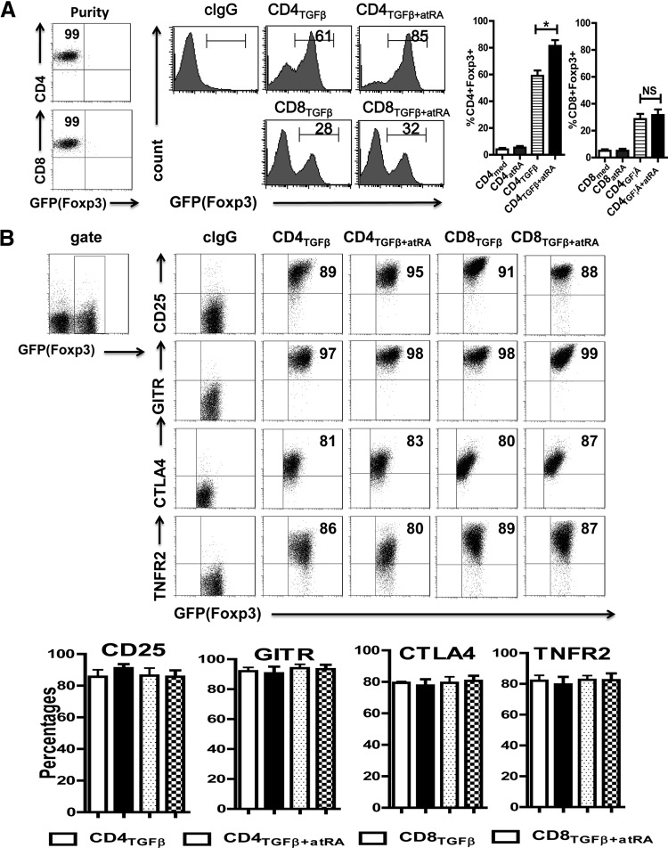
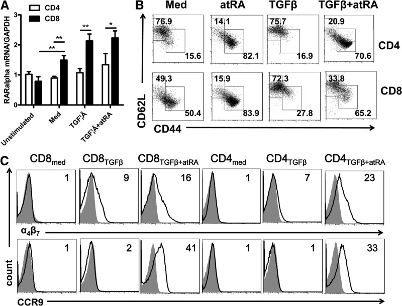
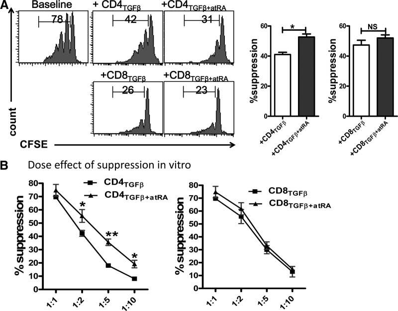
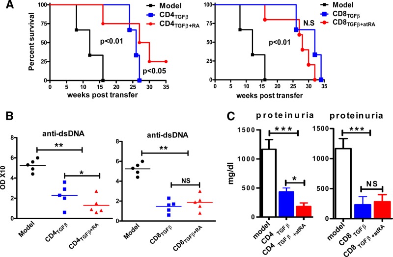
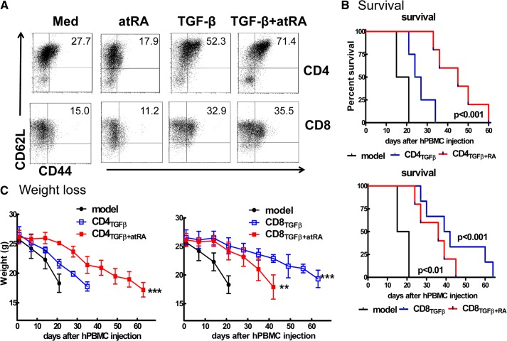
It is known that ATRA promotes the development of TGF-β-induced CD4(+)Foxp3(+) iTregs, which play a vital role in the prevention of autoimmune diseases; however, the role of ATRA in facilitating the differentiation and function of CD8(+)Foxp3(+) iTregs remains elusive. Using a head-to-head comparison, we found that ATRA promoted expression of Foxp3 and development of CD4(+) iTregs, but it did not promote Foxp3 expression on CD8(+) cells. Using a standard in vitro assay, we demonstrated that CD8(+) iTregs induced by TGF-β and ATRA were not superior to CD8(+) iTregs induced by TGF-β alone. In cGVHD, in a typical lupus syndrome model where DBA2 spleen cells were transferred to DBA2xC57BL/6 F1 mice, we observed that both CD8(+) iTregs induced by TGF-β and ATRA and those induced by TGF-β alone had similar therapeutic effects. ATRA did not boost but, conversely, impaired the differentiation and function of human CD8(+) iTregs. CD8(+) cells expressed the ATRA receptor RAR and responded to ATRA, similar to CD4(+) cells. We have identified the differential role of ATRA in promoting Foxp3(+) Tregs in CD4(+) and CD8(+) cell populations. These results will help to determine a protocol for developing different Treg cell populations and may provide novel insights into clinical cell therapy for patients with autoimmune diseases and those needing organ transplantation.

Keywords: Autoimmunity; Foxp3; GVHD; TGF-β; all-trans retinoic acid; regulatory T cells.


Figures

Figure 1.
Figure 1.. ATRA increased the percentages of Foxp3 expression on TGF-β-primed CD4+, but not on CD8+ cells.
(A) CD8+CD62L+CD25Foxp3(GFP) and CD4+CD62L+CD25Foxp3(GFP) cells isolated from C57BL/6 Foxp3gfp reporter mice were stimulated with immobilized anti-CD3 (1 μg/ml), soluble anti-CD28 (1 μg/ml), IL-2 (100 U/ml), or TGF-β (2 ng/ml), with (CD4TGFβ+ATRA or CD8TGFβ+ATRA) or without ATRA (50 nM) (CD4TGFβ or CD8TGFβ) for 3 days. Foxp3 (GFP) expression was examined by flow cytometry. Left: typical FACS histograms. Right: summary of data showing the frequency of Foxp3+ cells from TGF-β-primed CD4+ or CD8+ cells. *P < 0.05, NS. (B) The expression levels of regulatory T-cell associated markers including CD25, GITR, CTLA-4, and TNFR2 on CD4TGFβ, CD8TGFβ, CD4TGFβ+ATRA, or CD8TGFβ+ATRA cells were analyzed by flow cytometry. The graph data indicate the mean ± sem of 3 separate experiments showing the frequency of the indicated markers gated on the CD4 or CD8 cell populations.
Figure 2.
Figure 2.. CD8+ cells had the same response to ATRA as did CD4+ cells.
(A) The expression of ATRA RARα mRNA was determined by quantitative RT-PCR on naive or activated CD4+ and CD8+ cells. These cells were activated with TCR and IL-2, with or without TGF-β and with or without ATRA. Data are the mean ± se of 3 separate experiments. (B) CD4TGFβ, CD8TGFβ, CD4TGFβ+ATRA, or CD8TGFβ+ATRA cells were induced as in Fig. 1A, and the expression of CD44 and CD62L gated on CD4 or CD8 was analyzed by flow cytometry. Dot plots are representative of 3 independent experiments. (C) The expression of α4β7 and CCR-9 on CD4med, CD8med, CD4TGFβ, CD8TGFβ, CD4TGFβ+ATRA, or CD8TGFβ+ATRA cells was analyzed by flow cytometry. Data are representative of 3 independent experiments showing staining (black) plus isotype control (gray).
Figure 3.
Figure 3.. ATRA enhanced the suppressive activity of CD4+ iTregs, but not of CD8+ iTregs, in vitro.
(A) CD4TGFβ, CD8TGFβ, CD4TGFβ+ATRA, or CD8TGFβ+ATRA cells generated as in Fig. 1A were added to cultures of soluble anti-CD3 (0.025 μg/ml), irradiated APCs (1:1), and CFSE-labeled responder T cells for 3 days. Suppressive activity by each cell population was demonstrated and compared by percentages of CFSE-diluted cells. A histogram representative of 3 separate experiments in 1:2 ratios of Treg-to-responder T cells is shown. (B) The same culture as in (A), but the graded ratio of Tregs was added to the culture. The graph shows the mean ± sem of results in 3 independent experiments. *P < 0.05. **P < 0.01.
Figure 4.
Figure 4.. ATRA promoted the development of function of TGF-β-induced CD4+ iTregs, but not of CD8[supb]+ iTregs, in vivo.
CD4TGFβ, CD8TGFβ, CD4TGFβ+ATRA, or CD8TGFβ+ATRA cells were induced as in Fig. 1A. Freshly isolated splenocytes (80 × 106) from DBA/2 mice or together with 5 × 10⁶ of various CD4+ or CD8+ cell populations, as indicated, were intravenously transferred into C57BL/6xDBA/2 F1 mice. PBS was used for the negative control and marked as the model group. Each group had 6 mice, and the experiment was repeated twice. (A) Survival was monitored weekly. A Kaplan-Meier survival curve is shown. (B) The levels of anti-dsDNA IgG antibody in sera were measured by ELISA 4 weeks after cell transfer. (C) The proteinuria levels were examined 8 weeks after cell transfer. Data are combined from 2 independent experiments. *P < 0.05, **P < 0.01, ***P < 0.001.
Figure 5.
Figure 5.. ATRA promoted the function of TGF-β-induced human CD4+ iTregs, but not CD8+ iTregs, in a humanized xeno-GVHD model.
(A) Human CD4+CD45RA+ or CD8+CD45RA+ cells isolated from peripheral PBMCs were stimulated with anti-human CD3/CD28 beads 1:10 (1 bead:10 cells) with IL-2 (50–100 U/ml), with or without TGF-β1 (5 ng/ml) and with or without ATRA (100 nM) for 5 days. Foxp3 expression was determined by flow cytometry. Histogram data are representative of 3 independent experiments. (B) Kaplan-Meier survival estimates of NOD/SCID mice that received 20 × 106 CD25 hPBMCs or plus 5 × 10⁶ CD4TGFβ, CD8TGFβ, CD4TGFβ+ATRA, or CD8TGFβ+ATRA cells. (D) Body weight of xeno-GVHD model mice treated with or without conditioning cells, as described in (A). Data were collected from 3 independent experiments. *P < 0.05, **P < 0.01.

Similar articles

Cited by

References

    1. Sakaguchi S., Ono M., Setoguchi R., Yagi H., Hori S., Fehervari Z., Shimizu J., Takahashi T., Nomura T. (2006) Foxp3+ CD25+ CD4+ natural regulatory T cells in dominant self-tolerance and autoimmune disease. Immunol. Rev. 212, 8–27 - PubMed
    1. Horwitz D. A., Zheng S. G., Gray J. D. (2008) Natural and TGF-beta-induced Foxp3(+)CD4(+) CD25(+) regulatory T cells are not mirror images of each other. Trends Immunol. 29, 429–435 - PubMed
    1. Lan Q., Fan H., Quesniaux V., Ryffel B., Liu Z., Zheng S. G. (2012) Induced Foxp3(+) regulatory T cells: a potential new weapon to treat autoimmune and inflammatory diseases? J. Mol. Cell Biol. 4, 22–28 - PMC - PubMed
    1. Zhou X., Kong N., Zou H., Brand D., Li X., Liu Z., Zheng S. G. (2011) Therapeutic potential of TGF-beta-induced CD4(+) Foxp3(+) regulatory T cells in autoimmune diseases. Autoimmunity 44, 43–50 - PMC - PubMed
    1. Cosmi L., Liotta F., Lazzeri E., Francalanci M., Angeli R., Mazzinghi B., Santarlasci V., Manetti R., Vanini V., Romagnani P., Maggi E., Romagnani S., Annunziato F. (2003) Human CD8+CD25+ thymocytes share phenotypic and functional features with CD4+CD25+ regulatory thymocytes. Blood 102, 4107–4114 - PubMed

Publication types

MeSH terms