. 2013 Nov;163(3):1179-90.
doi: 10.1104/pp.113.223966. Epub 2013 Sep 30.

The nodulation factor hydrolase of Medicago truncatula: characterization of an enzyme specifically cleaving rhizobial nodulation signals

Affiliations

The nodulation factor hydrolase of Medicago truncatula: characterization of an enzyme specifically cleaving rhizobial nodulation signals

Ye Tian et al. Plant Physiol. 2013 Nov.

Abstract

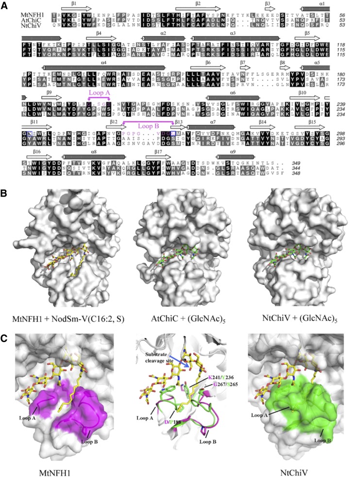
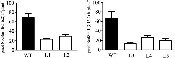
Nodule formation induced by nitrogen-fixing rhizobia depends on bacterial nodulation factors (NFs), modified chitin oligosaccharides with a fatty acid moiety. Certain NFs can be cleaved and inactivated by plant chitinases. However, the most abundant NF of Sinorhizobium meliloti, an O-acetylated and sulfated tetramer, is resistant to hydrolysis by all plant chitinases tested so far. Nevertheless, this NF is rapidly degraded in the host rhizosphere. Here, we identify and characterize MtNFH1 (for Medicago truncatula Nod factor hydrolase 1), a legume enzyme structurally related to defense-related class V chitinases (glycoside hydrolase family 18). MtNFH1 lacks chitinase activity but efficiently hydrolyzes all tested NFs of S. meliloti. The enzyme shows a high cleavage preference, releasing exclusively lipodisaccharides from NFs. Substrate specificity and kinetic properties of MtNFH1 were compared with those of class V chitinases from Arabidopsis (Arabidopsis thaliana) and tobacco (Nicotiana tabacum), which cannot hydrolyze tetrameric NFs of S. meliloti. The Michaelis-Menten constants of MtNFH1 for NFs are in the micromolar concentration range, whereas nonmodified chitin oligosaccharides represent neither substrates nor inhibitors for MtNFH1. The three-dimensional structure of MtNFH1 was modeled on the basis of the known structure of class V chitinases. Docking simulation of NFs to MtNFH1 predicted a distinct binding cleft for the fatty acid moiety, which is absent in the class V chitinases. Point mutation analysis confirmed the modeled NF-MtNFH1 interaction. Silencing of MtNFH1 by RNA interference resulted in reduced NF degradation in the rhizosphere of M. truncatula. In conclusion, we have found a novel legume hydrolase that specifically inactivates NFs.


Figures

Figure 1.
Figure 1.
Purification of recombinant proteins and activity test with NF substrates. A, SDS-PAGE analysis (left) and immunoblot detection (right) of His-tagged MtNFH1, Mt75352, MtCRA, AtChiC, and NtChiV expressed in E. coli BL21 (DE3). Purified proteins (1 μg) were analyzed on SDS-PAGE gels stained with Coomassie Brilliant Blue R-250. Immunoblot analysis was performed with a rabbit serum raised against MtNFH1 and the 3,3′-diamino-benzidine reagent. MW, Molecular mass. B, Separation of purified NFs and acylated cleavage products on a Nova Pak C18 column. The NF substrates were incubated with the indicated proteins at 37°C, extracted with n-butanol, and subjected to reverse-phase HPLC analysis. The cleavage products were separated into anomers (double peaks). Chromatograms are as follows: MtNFH1 (0.9 μg mL−1) incubated with 40 μm NodSm-V(C16:2, S) in 0.1 mL for 20 min (a), with 40 μm NodSm-IV(C16:2, S) in 0.1 mL for 40 min (b), with 20 μm desulfated NodSm-IV(C16:2) in 0.1 mL for 120 min (c), with 60 μm NodSm-IV(C16:2, Ac, S) in 0.1 mL for 40 min (d); the mutant protein MtNFH1(D148A) (0.9 μg mL−1) incubated with 20 μm NodSm-V(C16:2, S) in 0.1 mL for 4 h (e); Mt75352 (0.9 μg mL−1) incubated with 20 μm NodSm-V(C16:2, S) in 0.1 mL for 4 h (f); MtCRA (3 μg mL−1) incubated with 20 μm NodSm-V(C16:2, S) in 0.1 mL for 4 h (g); AtChiC (3 μg mL−1) incubated with 40 μm NodSm-V(C16:2, S) in 0.1 mL for 2 h (h); NtChiV (3 μg mL−1) incubated with 40 μm NodSm-V(C16:2, S) in 0.1 mL for 2 h (i); and MtNFH1 (0.9 μg mL−1) incubated with 20 μm NodSm-III(C16:2, S) in 0.1 mL for 4 h (j); II, NodSm-II(C16:2); II(Ac), NodSm-II(C16:2, Ac); III, NodSm-III(C16:2); IV, NodSm-IV(C16:2); IV(S), NodSm-IV(C16:2, S); IV(Ac, S), NodSm-IV(C16:2, Ac, S); V(S), NodSm-V(C16:2, S).
Figure 2.
Figure 2.
MtNFH1 does not hydrolyze nonmodified chitin oligosaccharides. Examples of HPLC chromatograms showing separation of the substrate (GlcNAc)6 and cleavage products are presented. After incubation (37°C) with the indicated proteins, reaction mixtures (in 25 mm sodium acetate buffer, pH 5.0; 30-μL test volume) were diluted with an equal volume of 50% acetonitrile and directly loaded onto the amino column. A, (GlcNAc)n standards (n = 1–6; 10 nmol). B, (GlcNAc)6 (5.8 mm) incubated with MtNFH1 (4.5 μg mL−1) for 3 h. C, (GlcNAc)6 (5.8 mm) incubated with AtChiC (5.0 μg mL−1) for 30 min. D, (GlcNAc)6 (5.8 mm) incubated with NtChiV (5.0 μg mL−1) for 30 min. I, GlcNAc; II, (GlcNAc)2; III, (GlcNAc)3; IV, (GlcNAc)4; V, (GlcNAc)5; VI, (GlcNAc)6; mAU, milliabsorbance units.
Figure 3.
Figure 3.
MtNFH1 does not inhibit the growth of the fungus T. viride. Purified proteins (3.2 μg in 50 μL) of NtChiV (1), AtChiC (2), or MtNFH1 (3) were pipetted into peripheral wells close to a growing mycelium of T. viride (inoculation at the center of the agar plate). The sodium acetate buffer (25 mm, pH 5.0) without enzyme was added as a control (4). The plate shown (demonstrating growth inhibition activity for NtChiV) was photographed after an incubation for 14 h at 27°C.
Figure 4.
Figure 4.
Homology modeling and docking simulation of NodSm-V(C16:2, S) to MtNFH1. A, Structure-based sequence alignment of MtNFH1 with AtChiC and NtChiV. Identical amino acid residues are shown on a black background, homologous residues on a gray background, and dashes indicate gaps; α-helices and β-strands are marked above the sequences. Amino acid residues of loops A and B in MtNFH1 are marked in magenta and the point-mutated residues K241 and G267 are framed in blue. B, Models of MtNFH1 with NodSm-V(C16:2, S), AtChiC with (GlcNAc)5, and NtChiV with (GlcNAc)5. C, MtNFH1 (magenta, left) has a prominent cleft between loops A and B that can bind the fatty acid moiety of NodSm-V(C16:2, S). NtChiV (green, right) and AtChiC lack such a binding cleft. The structures of MtNFH1 (magenta) and NtChiV (green) are superimposed to illustrate the expected conformational differences (middle). The cleavage site of NodSm-V(C16:2, S) hydrolyzed by MtNFH1 is indicated by a blue arrow.
Figure 5.
Figure 5.
Hydrolysis of NodSm-IV(C16:2, S) by roots of M. truncatula seedlings transformed with A. tumefaciens carrying pCAMBIA-MtNFH1i. Roots of seedlings (T2 generation) from five independent RNAi lines (named L1–L5) and wild-type plants (WT) were individually pretreated with 0.1 μm NodSm-IV(C16:2, S) for 1 d and then incubated with 15 μm NodSm-IV(C16:2, S) for 18 h. The NF substrate and formed NodSm-II(C16:2) were extracted from the medium with n-butanol and separated by reverse-phase HPLC using a Nova Pak C18 column. Data indicate means ± sd from two representative test series (in total, 24 transgenic and seven wild-type plants). NF-cleaving activities of all five RNAi lines are significantly reduced as compared with wild-type plants (Kruskal-Wallis test; P < 0.03).

References

    1. Altschul SF, Gish W, Miller W, Myers EW, Lipman DJ. (1990) Basic local alignment search tool. J Mol Biol 215: 403–410 - PubMed
    1. Antolín-Llovera M, Ried MK, Binder A, Parniske M. (2012) Receptor kinase signaling pathways in plant-microbe interactions. Annu Rev Phytopathol 50: 451–473 - PubMed
    1. Boller T, Gehri A, Mauch F, Vögeli U. (1983) Chitinase in bean leaves: induction by ethylene, purification, properties, and possible function. Planta 157: 22–31 - PubMed
    1. Bradford MM. (1976) A rapid and sensitive method for the quantitation of microgram quantities of protein utilizing the principle of protein-dye binding. Anal Biochem 72: 248–254 - PubMed
    1. Broghammer A, Krusell L, Blaise M, Sauer J, Sullivan JT, Maolanon N, Vinther M, Lorentzen A, Madsen EB, Jensen KJ, et al. (2012) Legume receptors perceive the rhizobial lipochitin oligosaccharide signal molecules by direct binding. Proc Natl Acad Sci USA 109: 13859–13864 - PMC - PubMed

Publication types

MeSH terms

Associated data