. 2013 Oct 15;110(42):16898-903.
doi: 10.1073/pnas.1304837110. Epub 2013 Sep 30.

Organ-specific function of adhesion G protein-coupled receptor GPR126 is domain-dependent

Affiliations

Organ-specific function of adhesion G protein-coupled receptor GPR126 is domain-dependent

Chinmoy Patra et al. Proc Natl Acad Sci U S A. .

Erratum in

  • Proc Natl Acad Sci U S A. 2014 Jan 21;111(3):1222

Abstract

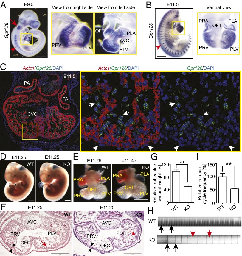
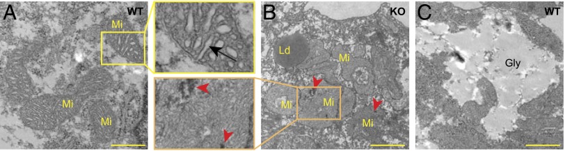
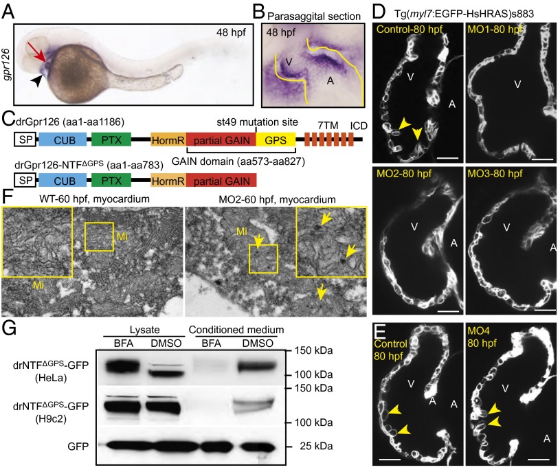
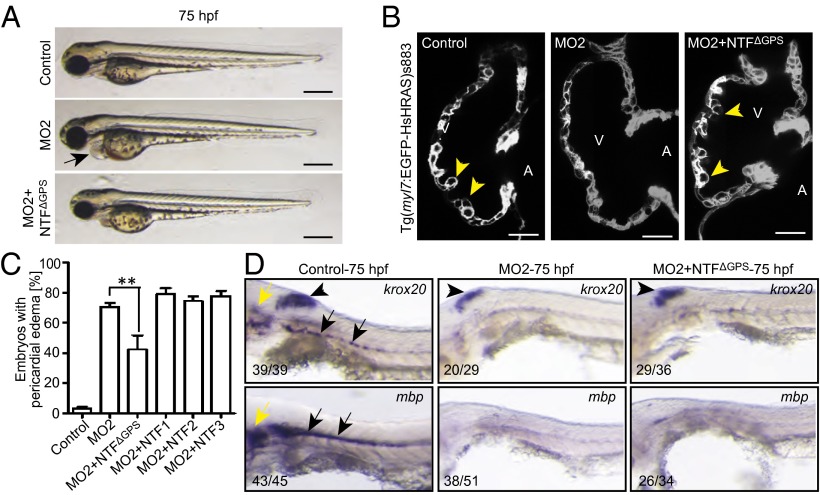
Despite their abundance and multiple functions in a variety of organ systems, the function and signaling mechanisms of adhesion G protein-coupled receptors (GPCRs) are poorly understood. Adhesion GPCRs possess large N termini containing various functional domains. In addition, many of them are autoproteolytically cleaved at their GPS sites into an N-terminal fragment (NTF) and C-terminal fragment. Here we demonstrate that Gpr126 is expressed in the endocardium during early mouse heart development. Gpr126 knockout in mice and knockdown in zebrafish caused hypotrabeculation and affected mitochondrial function. Ectopic expression of Gpr126-NTF that lacks the GPS motif (NTF(ΔGPS)) in zebrafish rescued the trabeculation but not the previously described myelination phenotype in the peripheral nervous system. These data support a model in which the NTF of Gpr126, in contrast to the C-terminal fragment, plays an important role in heart development. Collectively, our analysis provides a unique example of the versatile function and signaling properties of adhesion GPCRs in vertebrates.


Conflict of interest statement

The authors declare no conflict of interest.

Figures

Fig. 1.
Fig. 1.
Expression pattern of Gpr126 and characterization of R-Gpr126−/− mice. (A and B) Whole-mount in situ hybridization (lateral view) showing Gpr126 expression in the otic vesicle (red arrow), somites (red arrowhead), and heart (yellow square) at E9.5 in primitive ventricles [primitive left ventricle (PLV) and primitive right ventricle (PRV)], atrium [primitive right atrium (PRA) and primitive left atrium (PLA)], and the outflow tract (OFT) and at E11.5 in all primitive chambers but not in the OFT and interventricular region. (Scale bar: 500 µm.) (C) Multiplex in situ hybridization against alpha cardiac actin (Actc1; red) and Gpr126 (green) combined with DAPI staining (nuclei; blue) on thin sections indicating Gpr126 expression in endocardial (white arrows) but not epicardial cells (white arrowheads) and cardiomyocytes (red). (D and E) Gross morphology of WT and KO embryos (D) and hearts (E) at E11.25 is not different. (Scale bar: 500 µm.) (F) Hematoxylin and eosin staining of heart sections indicates hypotrabeculation (red arrows) and thinner ventricular myocardium (arrowheads) in KO hearts. (Scale bar: 300 µm.) (G) Quantification of the trabeculation phenotype. WT value was set to 100%. (H) Kymograph analysis. KO hearts displayed cardiac arrhythmia and bradycardia. Black lines/peaks (black arrows) represent ventricular contraction. Red arrows indicate irregular time intervals. (I) Quantitative analysis of H. WT value was set to 100%. Data are mean ± SEM. AVC, atrioventricular cushion; KO, knockout; OFC, outflow tract cushion; WT, wild type. **P < 0.01.
Fig. 2.
Fig. 2.
R-Gpr126−/− E11.25 cardiomyocytes contain defective mitochondria. (AC) Transmission electron micrographs displaying the ultrastructural details of cardiomyocytes in WT (A and C) and KO (B) hearts. (A) Mitochondria (Mi) with well-developed cristae (arrow) in WT cardiomyocytes. (B) KO cardiomyocytes contain abundant lipid droplets (Ld) and mitochondria with a more complex morphology, less well developed cristae, and electron-dense precipitates (arrowheads). (C) Glycogen (Gly) in WT cardiomyocytes. (Scale bar: 1 µm.)
Fig. 3.
Fig. 3.
Gpr126 knockdown leads to cardiac abnormalities in zebrafish. (A) Lateral view of a 48-hpf zebrafish embryo after whole-mount in situ hybridization showing gpr126 expression in heart (red arrow) and pericardium (black arrowhead). (B) Parasagittal section confirming cardiac expression of gpr126. (C) Schematic representation of zebrafish full-length Gpr126 (drGpr126) and its NTF part up to the st49 mutation site (drGpr126-NTFΔGPS). (D and E) Confocal sections of hearts from control- and morpholino-injected Tg(myl7:EGFP-HsHRAS)s883 embryos at 80 hpf. In full-length Gpr126-depleted animals (MO1-3) but not in Gpr126-CTF–depleted (MO4) morphants, trabeculation (yellow arrowheads) is perturbed. (Scale bar: 20 µm.) (F) Transmission electron micrographs at 60 hpf reveal that morphants contain elongated Mi with more branched cristae and electron dense precipitates (arrows) compared with WT siblings. (G) Western blot analysis of lysates and conditioned medium from cells overexpressing C-terminal GFP-tagged drGpr126-NTFΔGPS or GFP. Note that both GFP-tagged NTFΔGPS (predicted band size: 113 kDa) and GFP (predicted band size: 27 kDa) were detected in conditioned medium. However, secretion of GFP-tagged NTFΔGPS but not GFP was inhibited by BFA (a blocker of classical trans-Golgi secretory pathway). A, atrium; V, ventricle.
Fig. 4.
Fig. 4.
NTFΔGPS mRNA rescues the trabeculation phenotype in Gpr126-depleted zebrafish. (A) Lateral view of control-, MO2-, or MO2+NTFΔGPS mRNA-injected embryos at 75 hpf. MO2 injection resulted in pericardial edema (arrow). (B) Confocal sections of the hearts from control-, MO2-, and MO2+NTFΔGPS mRNA-injected Tg(myl7:EGFP-HsHRAS)s883 embryos at 80 hpf. In Gpr126-depleted animals, trabeculation (yellow arrowheads) is perturbed. Note that this phenotype is rescued by NTFΔGPS mRNA injection. (C) Quantitative analysis of rescue experiments of coinjections of MO2 and mRNAs encoding NTFΔGPS or NTF subfragments. Mean ± SEM. (D) Whole-mount in situ hybridization demonstrating krox20/egr2 and mbp expression in the otic vesicle (yellow arrow) and PLLn Schwann cells (black arrow) and krox20/egr2 expression in the hindbrain (arrowhead) at 75 hpf. In contrast, expression of krox20/egr2 and mbp is down-regulated in PLLn of MO2-mediated full-length Gpr126-depleted embryos. Coinjection of NTFΔGPS mRNA failed to rescue the PLLn myelineation phenotype in the cardiac phenotype-rescued animals. **P < 0.01.

References

    1. Bjarnadóttir TK, et al. The human and mouse repertoire of the adhesion family of G-protein-coupled receptors. Genomics. 2004;84(1):23–33. - PubMed
    1. Moriguchi T, et al. DREG, a developmentally regulated G protein-coupled receptor containing two conserved proteolytic cleavage sites. Genes Cells. 2004;9(6):549–560. - PubMed
    1. Prömel S, et al. The GPS motif is a molecular switch for bimodal activities of adhesion class G protein-coupled receptors. Cell Rep. 2012;2(2):321–331. - PMC - PubMed
    1. Kaur B, Brat DJ, Devi NS, Van Meir EG. Vasculostatin, a proteolytic fragment of brain angiogenesis inhibitor 1, is an antiangiogenic and antitumorigenic factor. Oncogene. 2005;24(22):3632–3642. - PubMed
    1. Koh JT, et al. Extracellular fragment of brain-specific angiogenesis inhibitor 1 suppresses endothelial cell proliferation by blocking alphavbeta5 integrin. Exp Cell Res. 2004;294(1):172–184. - PubMed

Publication types