Clinical Trial
. 2013 Oct 15;110(42):17053-8.
doi: 10.1073/pnas.1313650110. Epub 2013 Sep 30.

Paracrine activation of WNT/β-catenin pathway in uterine leiomyoma stem cells promotes tumor growth

Affiliations
Clinical Trial

Paracrine activation of WNT/β-catenin pathway in uterine leiomyoma stem cells promotes tumor growth

Masanori Ono et al. Proc Natl Acad Sci U S A. .

Abstract

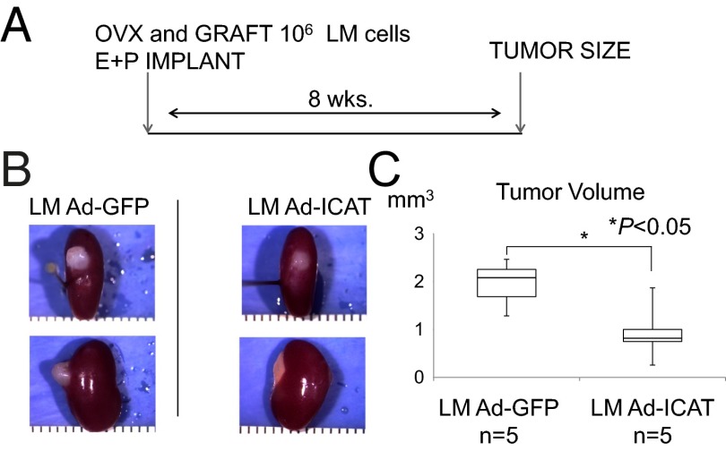
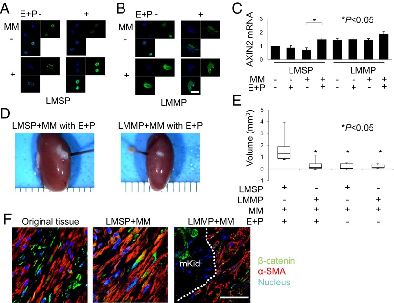
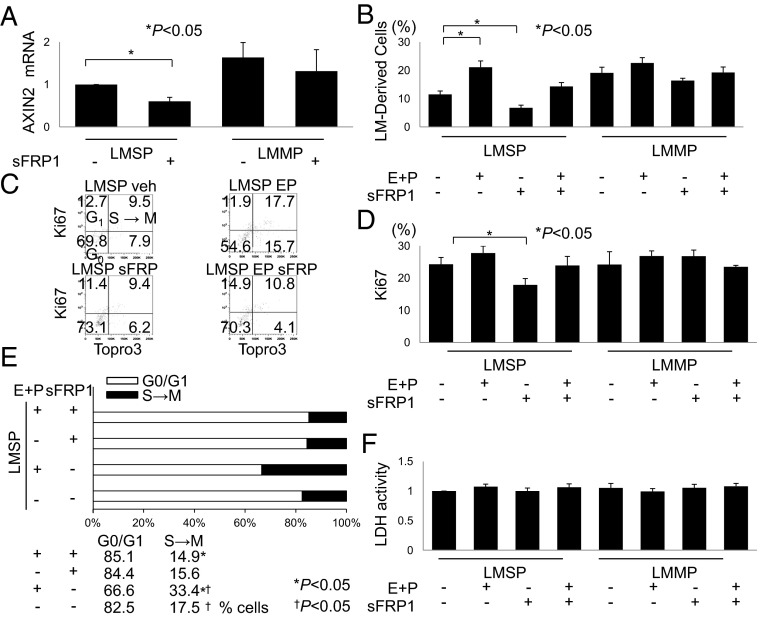
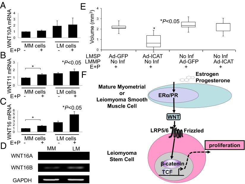
Uterine leiomyomas are extremely common estrogen and progesterone-dependent tumors of the myometrium and cause irregular uterine bleeding, severe anemia, and recurrent pregnancy loss in 15-30% of reproductive-age women. Each leiomyoma is thought to arise from a single mutated myometrial smooth muscle stem cell. Leiomyoma side-population (LMSP) cells comprising 1% of all tumor cells and displaying tumor-initiating stem cell characteristics are essential for estrogen- and progesterone-dependent in vivo growth of tumors, although they have remarkably lower estrogen/progesterone receptor levels than mature myometrial or leiomyoma cells. However, how estrogen/progesterone regulates the growth of LMSP cells via mature neighboring cells is unknown. Here, we demonstrate a critical paracrine role of the wingless-type (WNT)/β-catenin pathway in estrogen/progesterone-dependent tumorigenesis, involving LMSP and differentiated myometrial or leiomyoma cells. Estrogen/progesterone treatment of mature myometrial cells induced expression of WNT11 and WNT16, which remained constitutively elevated in leiomyoma tissues. In LMSP cells cocultured with mature myometrial cells, estrogen-progesterone selectively induced nuclear translocation of β-catenin and induced transcriptional activity of its heterodimeric partner T-cell factor and their target gene AXIN2, leading to the proliferation of LMSP cells. This effect could be blocked by a WNT antagonist. Ectopic expression of inhibitor of β-catenin and T-cell factor 4 in LMSP cells, but not in mature leiomyoma cells, blocked the estrogen/progesterone-dependent growth of human tumors in vivo. We uncovered a paracrine role of the WNT/β-catenin pathway that enables mature myometrial or leiomyoma cells to send mitogenic signals to neighboring tissue stem cells in response to estrogen and progesterone, leading to the growth of uterine leiomyomas.

Keywords: WNT/β-catenin signaling; paracrine signaling; tumor biology.


Conflict of interest statement

The authors declare no conflict of interest.

Figures

Fig. 1.
Fig. 1.
Blocking WNT activity inhibits the growth of LM tumors in NOD-SCID (IL2Rγnull) mice. (A) Experimental design and hormone treatment. Mice were ovariectomized and xenotransplanted with LM cells infected by Ad-GFP or Ad-ICAT and then were treated with s.c. implantation of E2 and P4 pellets. The mice underwent nephrectomy 8 wk after xenotransplantation. (B) Macroscopic visualization of the transplanted site 8 wk after xenotransplantation. (C) Xenografts were analyzed in terms of tumor volume. Error bars indicate SD; *P < 0.05; n = 5 paired patient samples.
Fig. 2.
Fig. 2.
E+P indirectly increases WNT signaling activity in LMSP cells. (A and B) Subcellular localization of β-catenin in LMSP or LMMP cells cultured in Transwells with or without MM coculture and in the presence or absence of E+P treatment. Indirect immunofluorescence was performed on LMSP and LMMP cells using anti–β-catenin antibody (1:100; green), followed by Hoechst nuclear counterstaining (blue). The overlay of β-catenin (green) and Hoechst (blue) is shown also. (Scale bar, 20 µm.) (C) Total RNA was extracted from LMSP or LMMP cells cultured in Transwells with or without MM to quantify the expression of AXIN2 by real-time quantitative PCR. Results are expressed as the mean ± SD of AXIN2 relative to GAPDH from four independent experiments. *P < 0.05. (D) Freshly isolated LMSP cells mixed with freshly isolated MM cells are able to generate larger LM tumors than mixes of LMMP-MM cells, with tumor sizes dependent on E+P treatment. Macroscopic visualization of the transplanted site 8 wk after xenotransplantation is shown. (E) Tumor volume of xenografts after 8 wk. Data are shown as mean ± SD. *P < 0.05. (F) Original tissue and generated tumor were analyzed by immunofluorescence. Immunostaining was performed to confirm the presence of β-catenin and α-SMA. Nuclei were stained with DAPI (blue). mKid, mouse kidney. Dotted line indicates the border between mouse kidney and tumor. (Scale bar, 30 µm.)
Fig. 3.
Fig. 3.
Inhibition of WNT signaling suppresses and E+P enhances the growth of LMSP cells cocultured with MM cells. (A) Effect of WNT signaling inhibition on LMSP-MM cocultured cells was determined by quantitative PCR of the WNT target gene AXIN2. Cells were seeded for mixed coculture as described previously and were treated with vehicle or sFRP1 (10 μg/mL) for 7 d. (B) Percentages of LMSP and LMMP cells among total cells were counted using PKH-26 dye and flow cytometry. Cells were seeded for mixed coculture and subjected to treatment with vehicle, 10−7 mol/L E+P, or sFRP1 (10 μg/mL) either alone or in combination with E+P (10−7 mol/L) for 7 d. Data are reported as the means ± SD. *P < 0.05. (CE) Cell-cycle analyses of LMSP and LMMP cells were performed using flow cytometry with the Ki-67 proliferation marker and DNA-specific labeling with the TO-PRO III fluorochrome. The percentages of positive or negative cells for both markers are indicated in each quadrant of the dot plots. The percentage of cells positive for the Ki-67 antigen was decreased after incubation with the WNT blocker sFRP1. Incubation with E+P increased the percentage of cells at the S→M phase compared with vehicle treatment, whereas incubation with E+P and sFRP did not increase the percentage of S→M phase cells. n = 3. *P < 0.05; P < 0.05. (F) LDH cell-viability assay. LMSP and LMMP cells were cultured with or without E+P and/or sFRP1 for 72 h, and media were assessed for LDH activity as described in SI Materials and Methods. Neither E+P treatment nor inhibition of WNT/β-catenin signaling promoted cell death. Results are expressed as levels of LDH activity (mean ± SD) from five independent experiments.
Fig. 4.
Fig. 4.
E+P treatment alters the expression of WNT genes in MM cells. (AC) MM and LM cells were treated with E+P or vehicle before isolation of RNA and evaluation of WNT expression by real-time quantitative PCR as described in SI Materials and Methods. Results represent the mean of a minimum of four independent experiments. Error bars indicate ± SD. *P < 0.05. (D) RT-PCR of WNT16B mRNA expression in MM and LM cells. GAPDH was used as a housekeeping gene control. (E) Ectopic ICAT expression in LMSP cells severely inhibited E+P-dependent tumor growth. *P < 0.05. (F) Interactions between ovarian steroids and WNT/β-catenin in LM tissue. Because estrogen receptor α (ERα) and progesterone receptor (PR) levels are remarkably higher in mature MM or LM cells than in stem cells, estrogen and progesterone likely affect LM stem cells in a paracrine fashion via estrogen receptor α and progesterone receptor expressed on adjacent mature MM or LM cells. In this model, estrogen and progesterone increase the secretion of WNT ligands from mature smooth muscle cells surrounding the stem cells. WNT via its FZD receptor then activates the β-catenin/TCF pathway in LM stem cells, inducing β-catenin target gene expression, cell proliferation, and tumorigenesis.

References

    1. Walker CL, Stewart EA. Uterine fibroids: The elephant in the room. Science. 2005;308(5728):1589–1592. - PubMed
    1. Wallach EE, Vlahos NF. Uterine myomas: An overview of development, clinical features, and management. Obstet Gynecol. 2004;104(2):393–406. - PubMed
    1. Okolo S. Incidence, aetiology and epidemiology of uterine fibroids. Best Pract Res Clin Obstet Gynaecol. 2008;22(4):571–588. - PubMed
    1. Stewart EA. Uterine fibroids. Lancet. 2001;357(9252):293–298. - PubMed
    1. Szotek PP, et al. Adult mouse myometrial label-retaining cells divide in response to gonadotropin stimulation. Stem Cells. 2007;25(5):1317–1325. - PubMed

Publication types

MeSH terms