

"Pseudo" Nomenclature in Dermatology: What's in a Name?
- PMID: 24082182
- PMCID: PMC3778777
- DOI: 10.4103/0019-5154.117305
"Pseudo" Nomenclature in Dermatology: What's in a Name?
Abstract
In the bewildering array of scientific nomenclature in the medical field, it is important to use correct terminology, know their aberrations and the reason behind a specific terminology. This paper is an attempt towards compiling all the pseudo-nomenclatures coined in dermatology, in order to make it easier to retain and recollect these pseudo names, signs, morphology, diseases, and conditions. It is also imperative to know the true entities that these pseudo names masquerade as, so as to understand the explanation for assigning the term 'pseudo' to these conditions. A total of 52 pseudo-terms have been compiled here in reference to dermatology. Most of these pseudo-nomenclatures were coined due to some clinical or histopathological resemblance to the true conditions, while some were premature conclusions drawn from a flawed understanding of the basic nature of the condition. Clear understanding of each of these terms and the explanation behind them being pseudo will enable a dermatologist to avoid misdiagnosis and needless confusion.
Keywords: Pseudo epitheliomatous hyperplasia; pseudo scleroderma; pseudo-Nikolskiy's sign.
Conflict of interest statement
Figures
References
-
- Valia AR, Ramesh V, Jerajani HR, Fernandez RJ. Blistering Disorders. In: Valia RG, Valia AR, editors. IADVL Textbook of dermatology. 3rd ed. Mumbai: Bhalani Publishing House; 2001. pp. 1087–152.
-
- Boyd AS, Nelder KH. The isomorphic response of Koebner. Int J Dermatol. 1990;29:401–10. - PubMed
-
- Stokes JH. Nevus pilaris with hyperplasia of non striated muscle. Arch Dermatol Syphil. 1923;7:479.
-
- Patton T, Zirwas M, Nieland-Fisher N, Jukic D. Inflammation of seborrheic keratoses caused by cytarabine: A pseudo sign of Leser-Trelat. J Drugs Dermatol. 2004;3:565–6. - PubMed
-
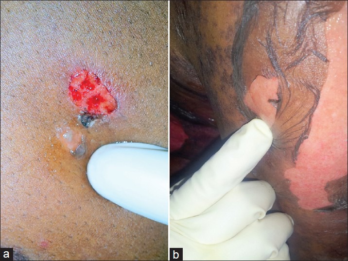
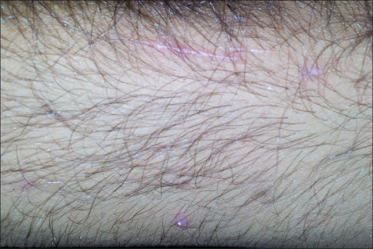
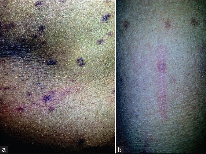
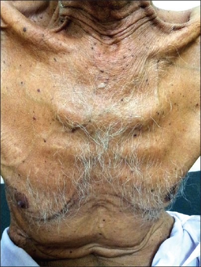
- Colomb D. Stellate spontaneous pseudoscars. Senile and presenile forms: Especially those forms caused by prolonged corticoid therapy. Arch Dermatol. 1972;105:551–4. - PubMed
LinkOut - more resources
Full Text Sources
Other Literature Sources