Rapid growth of left atrial myxoma after radiofrequency ablation
- PMID: 24082379
- PMCID: PMC3783123
Rapid growth of left atrial myxoma after radiofrequency ablation
Abstract
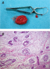
Atrial myxoma is the most common benign tumor of the heart, but its appearance after radiofrequency ablation is very rare. We report a case in which an asymptomatic, rapidly growing cardiac myxoma arose in the left atrium after radiofrequency ablation. Two months after the procedure, cardiovascular magnetic resonance, performed to evaluate the right ventricular anatomy, revealed a 10 × 10-mm mass (assumed to be a thrombus) attached to the patient's left atrial septum. Three months later, transthoracic echocardiography revealed a larger mass, and the patient was diagnosed with myxoma. Two days later, a 20 × 20-mm myxoma weighing 37 g was excised. To our knowledge, the appearance of an atrial myxoma after radiofrequency ablation has been reported only once before. Whether tumor development is related to such ablation or is merely a coincidence is uncertain, but myxomas have developed after other instances of cardiac trauma.
Keywords: Atrial fibrillation/prevention & control; catheter ablation/adverse effects; diagnosis, differential; echocardiography, transesophageal; echocardiography, transthoracic; heart neoplasms/ultrasonography; myxoma/diagnosis/surgery; pulmonary veins.
Figures


References
-
- Belhassen B, Rogowski O, Glick A, Viskin S, IIan M, Rosso R, Eldar M. Radiofrequency ablation of accessory pathways: a 14 year experience at the Tel Aviv Medical Center in 508 patients. Isr Med Assoc J 2007;9(4):265–70. - PubMed
-
- Abhishek F, Heist EK, Barrett C, Danik S, Blendea D, Correnti C, et al. Effectiveness of a strategy to reduce major vascular complications from catheter ablation of atrial fibrillation. J Interv Card Electrophysiol 2011;30(3):211–5. - PubMed
-
- Hahn K, Gal R, Sarnoski J, Kubota J, Schmidt DH, Bajwa TK. Transesophageal echocardiographically guided atrial transseptal catheterization in patients with normal-sized atria: incidence of complications. Clin Cardiol 1995;18(4):217–20. - PubMed
-
- Dhawan S, Tak T. Left atrial mass: thrombus mimicking myxoma. Echocardiography 2004;21(7):621–3. - PubMed
-
- Zhang FX, Yang B, Chen HW, Ju WZ, Cao KJ, Chen ML. Myocardial injury resulting from radiofrequency catheter ablation: comparison of circumferential pulmonary vein isolation and complex fractionated atrial electrograms ablation. Chin Med J 2011;124(17):2674–7. - PubMed
Publication types
MeSH terms
LinkOut - more resources
Full Text Sources
Medical
