Primary spinal intradural extramedullary lymphoma causing cauda equina syndrome
- PMID: 24082685
- PMCID: PMC3777313
- DOI: 10.4103/0974-8237.116538
Primary spinal intradural extramedullary lymphoma causing cauda equina syndrome
Abstract
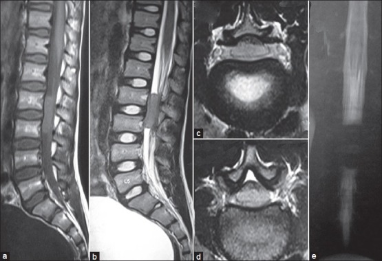
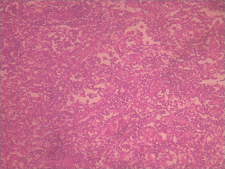
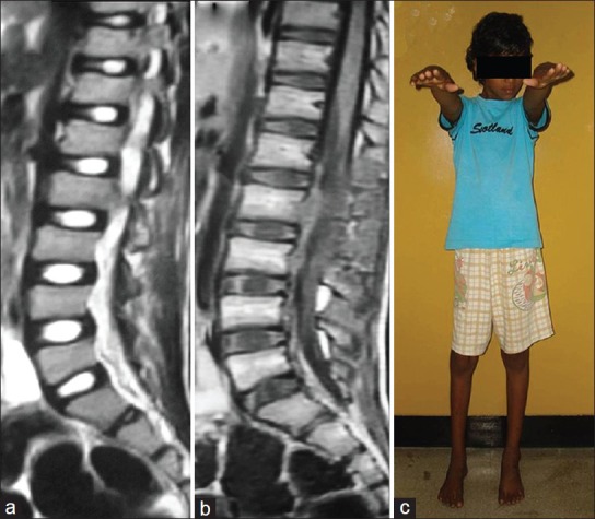
We report a case of lumbar intradural extramedullary lesion in an 11-year-old boy who presented with cauda equina syndrome and acute bladder disturbance. He underwent emergency surgical resection of the lesion, which was proved to be a lymphoma by histopathology and immunohistochemistry. He has improved neurologically and after 1 year, he is leading a normal life with near normal neurological functions. This is the second case of primary spinal intradural extramedullary lymphoma. This is the first such case in the pediatric age group and causing cauda equina syndrome. We describe the characteristics of such tumors along with pathogenesis and management.
Keywords: Cauda equina syndrome; chemoradiation; intradural extramedullary tumors; primary spinal lymphoma.
Conflict of interest statement
Figures
References
-
- Monnard V, Sun A, Epelbaum R, Poortmans P, Miller RC, Verschueren T, et al. Primary spinal epidural lymphoma: Patients’ profile, outcome, and prognostic factors: A multicenter Rare Cancer Network study. Int J Radiat Oncol Biol Phys. 2006;65:817–23. - PubMed
-
- Heran NS, Yong RL, Heran MS, Yip S, Fairholm D. Primary intradural extraarachnoid hodgkin lymphoma of the cervical spine. Case report. J Neurosurg Spine. 2006;5:61–4. - PubMed
-
- Yamashita T, Sakaura H, Oshima K, Iwasaki M, Yoshikawa H. Solitary intradural extramedullary lymphoma of the cervical spine. J Neurosurg Spine. 2010;12:436–9. - PubMed
-
- Marcotte P, Montpetit V, Burns B, Dennecy JM. Intradural spinal lymphoid hyperplasia. J Neuropathol Exp Neurol. 1990;49:261.
-
- Borovich B, Keret D, Ben-Arie J, Grushkiewicz I, Peyser E. Spinal intradural metastases of extraneural origin. Acta Neurochir (Wien) 1981;56:99–105. - PubMed