

Melanotic neuroectodermal tumor of infancy in the maxilla
- PMID: 24083035
- PMCID: PMC3780649
- DOI: 10.1155/2013/726815
Melanotic neuroectodermal tumor of infancy in the maxilla
Abstract
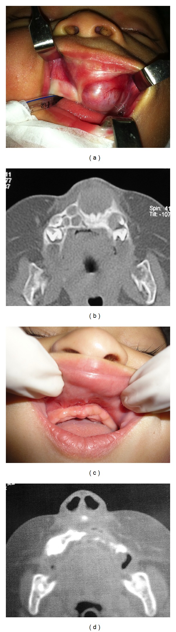
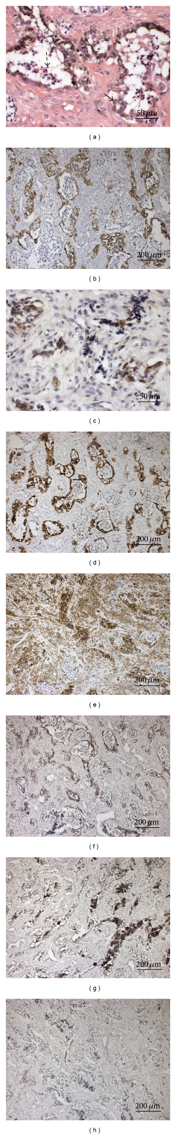
Melanotic neuroectodermal tumors of infancy (MNTIs) are rare fast-growing tumors with high recurrence rates. These tumors, which originate in the neural crest, commonly occur in the anterior maxilla of children under the age of one. Here, we describe an MNTI case in a two-month-old girl with increasing swelling in the left cheek. MNTI was diagnosed in this case following tomography and biopsy. The patient's histological and immunohistochemical profile indicated a remarkable combination of neural, melanocytic, and epithelial cell differentiation. One year following tumor excision, a follow-up examination revealed that the child exhibited no tumor recurrence. Approximately 260 cases of MNTI have been reported since this type of tumor was first described. In the present case, early diagnosis minimized the difficulties and risks associated with treatment and facilitated an optimal outcome. Despite complete surgical excision, careful followup is recommended. In addition, maxillary functional orthopedics and reconstruction may be necessary in cases of MNTI.
Figures
References
-
- Butt FMA, Guthua SW, Chindia ML, Rana F, Osundwa TM. Early outcome of three cases of melanotic neuroectodermal tumour of infancy. Journal of Cranio-Maxillofacial Surgery. 2009;37(8):434–437. - PubMed
-
- Barrett AW, Morgan M, Ramsay AD, Farthing PM, Newman L, Speight PM. A clinicopathologic and immunohistochemical analysis of melanotic neuroectodermal tumor of infancy. Oral Surgery, Oral Medicine, Oral Pathology, Oral Radiology, and Endodontics. 2002;93(6):688–698. - PubMed
-
- Chaudhary A, Wakhlu A, Mittal N, Misra S, Mehrotra D, Wakhlu AK. Melanotic neuroectodermal tumor of infancy: 2 decades of clinical experience with 18 patients. Journal of Oral and Maxillofacial Surgery. 2009;67(1):47–51. - PubMed
LinkOut - more resources
Full Text Sources
Other Literature Sources
