. 2013 Sep 13:2:460.
doi: 10.1186/2193-1801-2-460. eCollection 2013.

Immunohistochemical study of pituitary cells in wild and captive Salminus hilarii (Characiformes: Characidae) females during the annual reproductive cycle

Affiliations

Immunohistochemical study of pituitary cells in wild and captive Salminus hilarii (Characiformes: Characidae) females during the annual reproductive cycle

Renato Massaaki Honji et al. Springerplus. .

Abstract

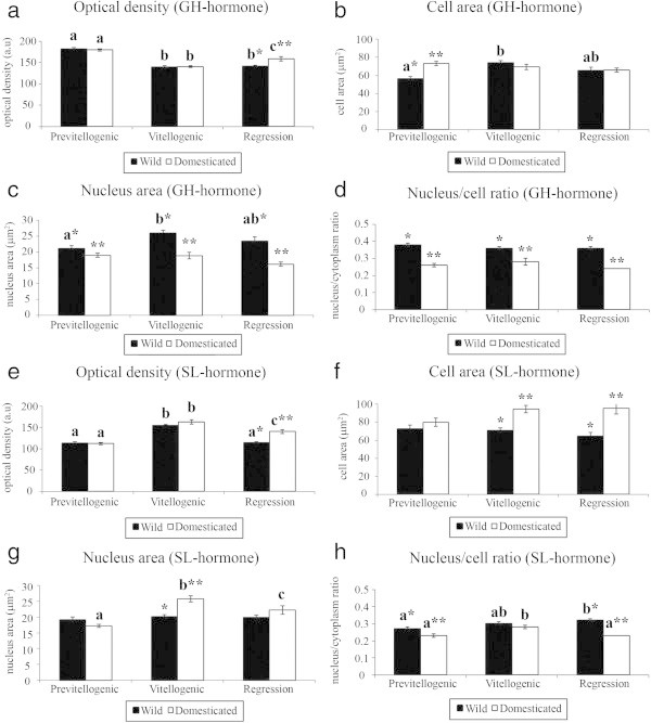
Freshwater fish that live exclusively in rivers are at particular risk from fragmentation of the aquatic system, mainly the species that migrate upriver for reproduction. That is the case of Salminus hilarii, an important migratory species currently classified as "almost threatened" in the São Paulo State (Brazil), facing water pollution, dam construction, riparian habitat destruction and environmental changes that are even more serious in this State. Additionally, this species show ovulation dysfunction in captivity. Our studies focused on the identification and distribution of the pituitary cell types in the adenohypophysis of S. hilarii females, including a morphometric analysis that compares pituitary cells from wild and captive broodstocks during the reproductive annual cycle. The morphology of adenohypophysial cells showed differences following the reproductive cycle and the environment. In general, optical density suggested a higher cellular activity during the previtellogenic (growth hormone) and vitellogenic (somatolactin) stages in both environments. Additionally, the nucleus/cell ratio analysis suggested that growth hormone and somatolactin cells were larger in wild than in captive females in most reproductive stages of the annual cycle. In contrast, prolactin hormone showed no variation throughout the reproductive cycle (in both environments). Morphometrical analyses related to reproduction of S. hilarii in different environmental conditions, suggest that somatolactin and growth hormone play an important role in reproduction in teleost and can be responsible for the regulation of associated processes that indirectly affect reproductive status.

Keywords: Gonadotropins; Growth hormone; Prolactin hormone; Reproductive dysfunction; Somatolactin hormone.


Figures

Figure 1
Figure 1
Environmental data obtained from the Brazilian Government Agencies. a) Water temperature (Upper Tietê River and Ponte Nova Fish Farm); b) pluviometric index; c) Photoperiod. Sources: “Departamento de Água e Energia Elétrica (DAEE)”, “Instituto Agronômico e Geofísico (IAG-USP), and “Companhia de Tecnologia de Saneamento Ambiental (CETESB)”.
Figure 2
Figure 2
Sagittal sections through the pituitary gland ofSalminus hilarii. a) Photomicrograph of the stained GH cells. Examples of measured nuclear area (red circle) and cell area (black circle) and the optical density (white square) were indicated; b-c) Sagittal sections of the pituitary stained using Mallory trichrome (b) and periodic acid-Schiff (PAS); d) Schematic sagittal representation of the pituitary, showing the distribution of ADH cells. RPD: rostral pars distalis; PPD: proximal pars distalis; PI: pars intermedia; NH: neurohypophysis; (■) prolactin cells; (●) growth hormone; (☆) gonadotropins cells; (▲) somatolactin cells; e) PRL-ir cells in the RPD. Details of PRL cells using the anti-chum salmon antisera (inset); f, g, h) GH-ir cells in the PPD (f) using the anti-chum salmon antisera. Details of cluster (arrowhead) of GH-ir cells (g) and isolated (arrowhead) of GH-ir cells (h). Scale bars: a, g-h) 20 μm; b-c, e-f) 300 μm (20 μm insert).
Figure 3
Figure 3
Sagittal sections through the pituitary gland ofSalminus hilarii. a-d) β-FSH and β-LH cells using the anti-chum salmon antiserum (Dr. H. Kawauchi). β-FSH-ir cells (a) and β-LH-ir cells (b) in the PPD showing the weakly immunoreactivity to the anti-salmon. Details of β-FSH cells immunoreactive (c) (arrowhead) corresponding to a high magnification of (a). Details of β-LH cells (d) (arrowhead) corresponding to a high magnification of (b); e-h) β-FSH and β-LH cells using the anti-chum salmon antiserum (Dr. A. Shimizu). β-FSH-ir cells (e) and β-LH-ir cells (f) in the PPD showing the weakly immunoreactivity to the anti-salmon. Details of β-FSH cells (g) (arrowhead) and β-LH cells (h) (arrowhead) corresponding to a high magnification of (e) and (f) respectively. For more information, see the explanation in the text. RPD: rostral pars distalis; PPD: proximal pars distalis; PI: pars intermedia. Scale bars: a, b, e, f) 300 μm; c, d, g, h) 50 μm.
Figure 4
Figure 4
Sagittal sections through the pituitary gland ofSalminus hilarii. a-d) Sections of pituitary showing the localization of α-β-GtHs and β-GtHs cells, using single immunocytochemistry, with the anti-carp antiserum (Dr. J. Mancera). a, b) Gonadotropins cells widely distributed in PPD; c, d) Details and differentiated GtH-ir cells (arrowhead) using anti-carp antisera, in a major magnitude of (a) and (b) respectively; e,f) Sections of the pituitary showing the localization of SL cells using single immunocytochemistry. Details of SL cells (arrowhead) (f) in high magnitfication of (e); g) periodic acid-Schiff (arrowheads indicate the PAS reactivity to SL cells); h) cross-reaction using SL antibody in GtH cells (arrowheads) in PPD region; i) PRL cells in RPD region with weakly cross-reaction, when using SL antibody. For more information, see the explanation in the text. RPD: rostral pars distalis; PPD: proximal pars distalis; PI: pars intermedia. Scale bars: a,b, e) 300 μm; c,d, i) 50 μm; f, g, h) 20 μm.
Figure 5
Figure 5
Salminus hilarii.Semiquantitative analysis of growth hormone (GH) and somatolactin hormone (SL) in the different environments and ovarian maturation stages. a) optical density of GH; b) cell area of GH; c) nucleus area of GH; d) nucleus/cell ratio of GH; e) optical density of SL; f) cell area of SL; g) nucleus area of SL; h) nucleus/cell ratio of SL. Values followed by different letters (a, b) are significantly different during the reproductive cycle. Values followed by different symbols (*) are significantly different between the environments. Each bar represents the mean ± SEM. Significance (P < 0.05).

Similar articles

Cited by

References

    1. Agostinho AA, Gomes LC, Suzuki HI, Júlio HF., Jr . Upper Paraná river basin Brazil. In: Carolsfeld J, Harvey B, Ross C, Baer A, editors. Migratory fishes of south America: biology, fisheries and conservation status. Victoria: World Fisheries Trust, The World Bank and the International Development Research Centre; 2003. pp. 19–98.
    1. Amaral JS, Melo RG, Honji RM, Moreira RG. Effects of migration impediment of Salminus hilarii (teleost: Characidae) on the pituitary-gonad axis. Comp Biochem Physiol A. 2007;148S:44.
    1. Andrade DR, Godinho AL, Godinho HP, Shimoda E. Biologia reprodutiva da tabarana, Salminus hilarii (Osteichthyes, Characidae) na represa de Três Marias, MG. Rev Bras Med Vet. 2006;28(1):26–32.
    1. Araújo BC, Mello PH, Honji RM, Moreira RG. The influence of captive breeding on the fatty acid profiles of Salminus hilarii (Characiformes: Characidae) eggs and larvae. Aquac Int. 2012;20:1161–1181. doi: 10.1007/s10499-011-9472-6. - DOI
    1. Batten TFC. Immunocytochemical demonstration of pituitary cell types in the teleost Poecilia latipinna, by light and electron microscopy. Gen Comp Endocrinol. 1986;63:139–154. doi: 10.1016/0016-6480(86)90192-9. - DOI - PubMed

LinkOut - more resources