Review
. 2013;14(9):213.
doi: 10.1186/gb-2013-14-9-213.

Neurexins

Review

Neurexins

Carsten Reissner et al. Genome Biol. 2013.

Abstract

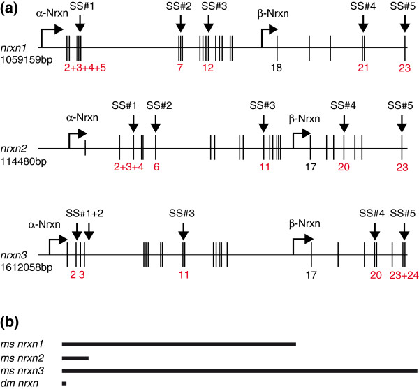
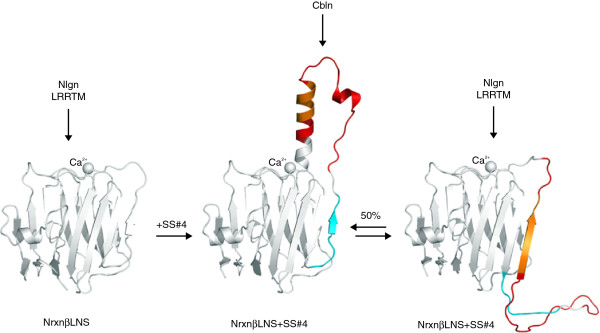
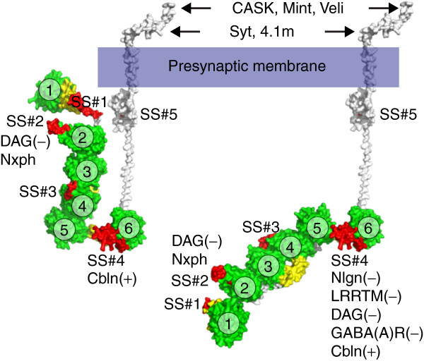
The neurexin family of cell adhesion proteins consists of three members in vertebrates and has homologs in several invertebrate species. In mammals, each neurexin gene encodes an α-neurexin in which the extracellular portion is long, and a β-neurexin in which the extracellular portion is short. As a result of alternative splicing, both major isoforms can be transcribed in many variants, contributing to distinct structural domains and variability. Neurexins act predominantly at the presynaptic terminal in neurons and play essential roles in neurotransmission and differentiation of synapses. Some of these functions require the formation of trans-synaptic complexes with postsynaptic proteins such as neuroligins, LRRTM proteins or cerebellin. In addition, rare mutations and copy-number variations of human neurexin genes have been linked to autism and schizophrenia, indicating that impairments of synaptic function sustained by neurexins and their binding partners maybe relevant to the pathomechanism of these debilitating diseases.


Figures

Figure 1
Figure 1
Domain organization of α-neurexins and β-neurexins. Neurexins are type I transmembrane proteins with a single path transmembrane helix (TM) that separates amino-terminal extracellular from cytosolic intracellular domains. The hallmark of neurexins is a cassette of LNS(green)-EGF(orange)-LNS(green) that is repeated three times in α-neurexin (Nrxn1α), albeit with low sequence conservation (16% identity and 27% homology). β-Neurexin (Nrxn1β) starts with its own exon that encodes a signal peptide (SP) and unique 37 histidine-rich residues (blue). The remainder is identical to the corresponding α-neurexin starting from the last LNS domain. Red symbols indicate positions of up to five canonically conserved splice sites (SS#1 to SS#5), and hexamers point to N-glycosylation sites and O-glycosylation sites. EGF, epidermal growth factor-like domain; LNS, laminin-neurexin-sex hormone binding globulin.
Figure 2
Figure 2
Genomic organization of neurexin genes. (a) Gene organization of mouse neurexins (nrxn) with exons (vertical lines) segregating introns (horizontal lines). The nrxn2 gene is 10 times smaller than nrxn1 or nrxn3 due to shorter introns but the relative positions of transcription starts for α-variants and β-variants (kinked arrows) are similar in all cases. Red numbers indicate alternatively spliced exons, while β-specific exons are in black. The first splice site (SS#1) accepts different inserts derived from combinations of two to four mini exons, whereas others such as SS#2 can also use parts of an insert sequence from one exon. (b) Vertebrate nrxn genes are up to 100 times longer than the single nrxn from invertebrates. The length ratio of Drosophila (dm nrxn) to mouse neurexins (ms nrxn) 2 and 3 is 1:10:100, respectively.
Figure 3
Figure 3
Phylogenetic tree of the neurexin protein family. Dendrogram showing the phylogenetic relationships between the vertebrate and invertebrate neurexins. The tree was generated using neurexin amino acid sequences from several vertebrate species and invertebrate homologs, and a gap-free sequence alignment with GeneBee [132]. The neurexin 1 (Nrxn1) family is shown in red, neurexin 2 (Nrxn2) in blue, and neurexin 3 (Nrxn3) in green. The invertebrate sequences are shown in black. Species names and GenBank accession numbers [133] are given for each branch. Cluster distance values indicated at branches represent the amino acid differences for the particular group of sequences. Note that the more distantly related Caspr/paranodin/CTNAP family member ‘neurexin 4’ contains a different domain structure and is not included in the analysis.
Figure 4
Figure 4
LNS domains as a versatile toolbox for protein-protein interactions. The diagram shows a ribbon structure of αLNS6 (PDB ID: 2R1D) representing the lowest common denominator of the six neurexin LNS folds; it is used here to highlight specific features among the individual domains. The fold is formed by 14 β-strands (β1 to 14), which are generally tightly connected. In αLNS6/βLNS, β10 (blue) can be displaced by an alternatively spliced insert at SS#4 (red). The synopsis also shows that positions of splice sites SS#2 (green) from αLNS2, SS#3 (orange) from αLNS4, and SS#4 from αLNS6 are all in vicinity of the corresponding Ca2+-binding site. The splice insert in SS#4 participates in Ca2+ coordination, while an insert in the SS#3 domain might prevent Ca2+ association in adjacent αLNS3. In the αLNS3 domain, the β4/β5 loop (magenta) is prolonged and can be interpreted as a permanent splice insert that interacts with the insert in SS#3. These β-loop variations individually shape each LNS domain around the Ca2+-binding site suitable to mediate specific LNS-protein or LNS-glycan interactions. LNS, laminin-neurexin-sex hormone binding globulin.
Figure 5
Figure 5
Splice insert in SS#4 causes a molecular switch. Splice insert-free βLNS-SS#4 (PDB ID: 3B3Q; left panel) can bind efficiently to neuroligin (Nlgn) and leucine-rich repeat proteins (LRRTM), which have overlapping binding epitopes. The prolonged conformation caused by an insert in SS#4 (orange/red; from PDB ID: 2R1B) blocks binding to Nlgn and LRRTM, and instead allows the binding of cerebellin (Cbln, middle panel). This structure of βLNS + SS#4 is in equilibrium with an additional conformation (PDB ID: 3 MW2), in which β10 (cyan) is replaced by part of the SS#4 insert (orange, right panel). In the latter, Nlgn and LRRTM binding is restored, while interaction with Cbln should be abolished. The diagrams were made using the actual structural coordinates and PyMOL software (Schrödinger, Mannheim, Germany).
Figure 6
Figure 6
Structural models of α-neurexin. The diagram visualizes conformations that the extracellular domain of α-neurexin can assume. In the U-form (modeled from PDB ID: 3R05; left) only cerebellin (Cbln), neurexophilin (Nxph) and dystroglycan (DAG) might bind to LNS6 and LNS2, respectively. After rotation of about 180° in the αLNS5-αLNS6 hinge (modeled using PDB ID: 3ASI and 3R05; right), the core structure and αLNS6 become elongated and accessible to additional ligands, including neuroligins (Nlgn) and leucine-rich repeat molecules (LRRTM). The parentheses indicate the required presence (+) or absence (−) of the splice inserts in αLNS6 (SS#4) or αLNS2 (SS#2). Coordinates for αLNS1 have been modeled by sequence homology to other LNS domains because its electron density map was not resolved in the crystal structure [60]. Intracellularly, cytosolic proteins such as synaptotagmin (Syt), protein 4.1 from brain (4.1 m), CASK, Mint and Veli bind to the disordered carboxy-terminal domain of neurexins. LNS domains, green (numbered 1 to 6); EGF-like domains, yellow; splice inserts at splice sites #1 to #5, red. EGF, epidermal growth factor-like; LNS, laminin-neurexin-sex hormone binding globulin.
Figure 7
Figure 7
Trans-synaptic neurexin-neuroligin complexes shape excitatory and inhibitory synapses. Presynaptic α-neurexins or β-neurexins (red) can interact with dimeric neuroligins (green) across the synaptic cleft to regulate important aspects of establishment, differentiation and maturation of synapses. Isoforms and splice variants of both molecules have been proposed to be differentially distributed at excitatory or inhibitory synapses to establish specificity. Note that presence of β-neurexins (β-Nrxn) at inhibitory terminals is unclear, while for neuroligins (Nlgn), Nlgn2 and Nlgn4 show quite specific localization and roles at inhibitory synapses. Intracellularly, the cytosolic domains of Nrxn and Nlgn are able to cluster components of the presynaptic release machinery and of postsynaptic signaling pathways and transmitter receptors (R). The clustering ability of Nrxn and Nlgn variants at excitatory or inhibitory synapses is mostly derived from cell culture assays. AMPAR, α-amino-3-hydroxy-5-methyl-4-isoxazolepropionic acid receptor; GABA, γ-aminobutyric acid; NMDAR, N-methyl-d-aspartate receptor; PSD95, postsynaptic density protein-95; VGat, vesicular GABA transporter; VGlu, vesicular glutamate transporter.

References

    1. Ullrich B, Ushkaryov YA, Südhof TC. Cartography of neurexins: more than 1000 isoforms generated by alternative splicing and expressed in distinct subsets of neurons. Neuron. 1995;14:497–507. - PubMed
    1. Ushkaryov YA, Hata Y, Ichtchenko K, Moomaw C, Afendis S, Slaughter CA, Südhof TC. Conserved domain structure of beta-neurexins. Unusual cleaved signal sequences in receptor-like neuronal cell-surface proteins. J Biol Chem. 1994;14:11987–11992. - PubMed
    1. Ushkaryov YA, Petrenko AG, Geppert M, Südhof TC. Neurexins: synaptic cell surface proteins related to the alpha-latrotoxin receptor and laminin. Science. 1992;14:50–56. - PubMed
    1. Etherton MR, Blaiss CA, Powell CM, Südhof TC. Mouse neurexin-1alpha deletion causes correlated electrophysiological and behavioral changes consistent with cognitive impairments. Proc Natl Acad Sci U S A. 2009;14:17998–18003. - PMC - PubMed
    1. Kattenstroth G, Tantalaki E, Südhof TC, Gottmann K, Missler M. Postsynaptic N-methyl-d-aspartate receptor function requires alpha-neurexins. Proc Natl Acad Sci U S A. 2004;14:2607–2612. - PMC - PubMed

Publication types

MeSH terms