. 2013 Oct 1;3(1):23.
doi: 10.1186/2044-5040-3-23.

Control of mRNA stability contributes to low levels of nuclear poly(A) binding protein 1 (PABPN1) in skeletal muscle

Affiliations

Control of mRNA stability contributes to low levels of nuclear poly(A) binding protein 1 (PABPN1) in skeletal muscle

Luciano H Apponi et al. Skelet Muscle. .

Abstract

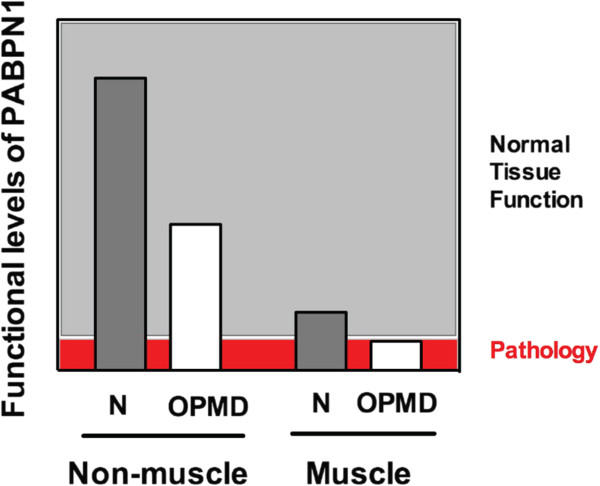
Background: The nuclear poly(A) binding protein 1 (PABPN1) is a ubiquitously expressed protein that plays critical roles at multiple steps in post-transcriptional regulation of gene expression. Short expansions of the polyalanine tract in the N-terminus of PABPN1 lead to oculopharyngeal muscular dystrophy (OPMD), which is an adult onset disease characterized by eyelid drooping, difficulty in swallowing, and weakness in the proximal limb muscles. Why alanine-expanded PABPN1 leads to muscle-specific pathology is unknown. Given the general function of PABPN1 in RNA metabolism, intrinsic characteristics of skeletal muscle may make this tissue susceptible to the effects of mutant PABPN1.

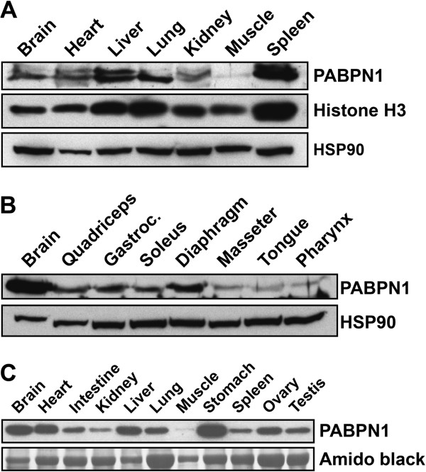
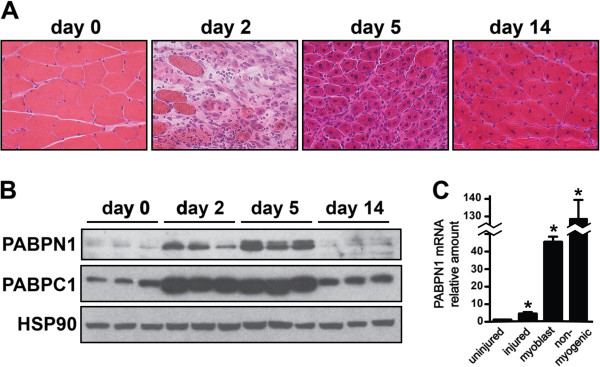
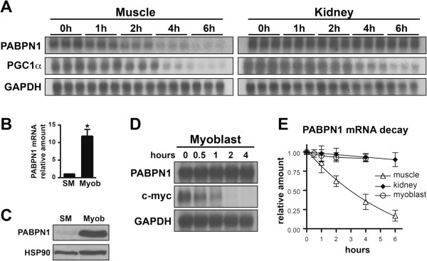
Methods: To begin to understand the muscle specificity of OPMD, we investigated the steady-state levels of PABPN1 in different tissues of humans and mice. Additionally, we analyzed the levels of PABPN1 during muscle regeneration after injury in mice. Furthermore, we assessed the dynamics of PABPN1 mRNA decay in skeletal muscle compared to kidney.

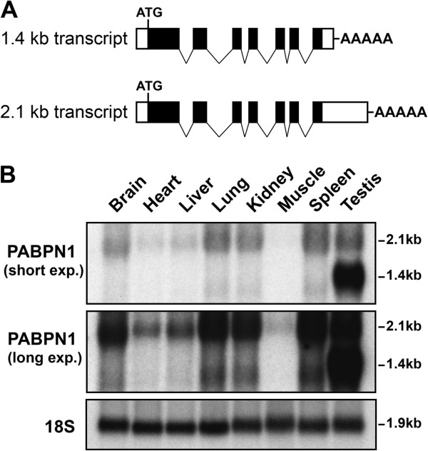
Results: Here, we show that the steady-state levels of both PABPN1 mRNA and protein are drastically lower in mouse and human skeletal muscle, particularly those impacted in OPMD, compared to other tissues. In contrast, PABPN1 levels are increased during muscle regeneration, suggesting a greater requirement for PABPN1 function during tissue repair. Further analysis indicates that modulation of PABPN1 expression is likely due to post-transcriptional mechanisms acting at the level of mRNA stability.

Conclusions: Our results demonstrate that PABPN1 steady-state levels and likely control of expression differ significantly in skeletal muscle as compared to other tissues, which could have important implications for understanding the muscle-specific nature of OPMD.


Figures

Figure 1
Figure 1
Nuclear poly(A) binding protein 1 (PABPN1) levels are low in all skeletal muscles. Lysates prepared from different (A) mouse tissues (50 μg of total protein per lane), (B) mouse muscles (150 μg of total protein per lane) or (C) human tissues (20 μg of total protein per lane) were immunoblotted with anti-PABPN1 antibody. Histone H3 and heat shock protein 90 (HSP90) were used as loading controls for mouse samples. Amido black staining was used as the loading control for human samples. Immunoblots are representative of at least three independent sets of tissues.
Figure 2
Figure 2
Nuclear poly(A) binding protein 1 (PABPN1) mRNA levels are low in skeletal muscle. (A) Structure of the 2.1 kb and 1.4 kb PABPN1 transcripts (solid boxes represent coding regions and open boxes non-coding regions). (B) Total RNA from different mouse tissues was analyzed by northern blot using a PABPN1 probe. Two different exposures, short and long, are shown. 18S rRNA was probed as a loading control. Figure is representative of at least three independent sets of tissues.
Figure 3
Figure 3
Nuclear poly(A) binding protein 1 (PABPN1) levels are increased during muscle regeneration in part due to increased levels in myoblasts. (A) Representative hematoxylin and eosin stained sections of gastrocnemius muscles at different times after BaCl2 injury are shown. (B) Lysates were prepared from gastrocnemius muscles at different times after injury and were immunoblotted with anti-PABPN1, PABPC1 or heat shock protein 90 (HSP90) antibodies (n = 3 per timepoint). (C) Total RNA was obtained from uninjured and injured muscle tissue (three independent samples) as well as fluorescence-activated cell sorting (FACS)-sorted myoblasts and non-myogenic cells obtained 3 days after muscle injury (pooled from five mice). PABPN1 mRNA levels were determined using real-time polymerase chain reaction (PCR) and hypoxanthine-guanine phosphoribosyltransferase (HPRT) mRNA was used as an internal control. Amount of PABPN1 mRNA relative to uninjured muscle is shown; n = 3. Data are mean ± SD; *P <0.05 vs uninjured muscle.
Figure 4
Figure 4
Nuclear poly(A) binding protein 1 (PABPN1) mRNA is unstable in muscle tissue but stable in cultured myoblasts. (A) Total RNA was collected at different timepoints after injection of actinomycin D to inhibit transcription and PABPN1 mRNA decay was analyzed by northern blot. Time courses are shown for samples from muscle and kidney. Peroxisome proliferator-activated receptor gamma coactivator 1α (PGC1α) and glyceraldehyde 3-phosphate dehydrogenase (GAPDH), a known unstable and stable transcript, respectively, were probed as controls (n = 3 per timepoint). To visualize PABPN1 signal in muscle samples, the blot was exposed significantly longer than for kidney samples. (B) Total RNA was obtained from skeletal muscle (SM) and cultured primary mouse myoblasts (Mb) and PABPN1 mRNA levels were determined using real-time polymerase chain reaction (PCR) and hypoxanthine-guanine phosphoribosyltransferase (HPRT) mRNA was used as an internal control. The amount of PABPN1 mRNA relative to skeletal muscle (SM) is shown; n = 3 independent samples. Data are mean ± SD; *P <0.05 vs skeletal muscle. (C) Protein extracts were prepared from SM and Mb and immunoblotted with anti-PABPN1 antibody. HSP90 was used as a loading control. The immunoblot is representative of at least three independent samples. (D) Total RNA was collected at different timepoints after treatment of cultured primary mouse myoblasts with actinomycin D and PABPN1 mRNA decay was analyzed by northern blot; c-Myc and GAPDH, known unstable and stable transcripts in myoblasts, respectively, were probed as controls. Averages of densitometric measurements of northern blot bands were used to determine mRNA decay. The image is representative of at least three independent samples. (E) The decay profile of PABPN1 mRNA in muscle, kidney and cultured myoblasts plotted as mRNA amount relative to timepoint T = 0 h (n = 3 samples per timepoint). Data are mean ± SD.
Figure 5
Figure 5
Low levels of nuclear poly(A) binding protein 1 (PABPN1) in skeletal muscle may predispose this tissue to the deleterious effects of alanine-expanded PABPN1. We show muscle has lower levels of PABPN1 compared to other tissues in normal individuals (N) but these levels are adequate for normal tissue function. In patients with oculopharyngeal muscular dystrophy (OPMD), functional levels of PABPN1 could be decreased in all tissues due to expression of mutant PABPN1. However, muscle-specific pathology ensues in autosomal dominant OPMD because the levels of PABPN1 fall below the threshold required to maintain proper tissue function.

References

    1. Keene JD. RNA regulons: coordination of post-transcriptional events. Nat Rev Genet. 2007;3:533–543. doi: 10.1038/nrg2111. - DOI - PubMed
    1. Kuhn U, Wahle E. Structure and function of poly(A) binding proteins. Biochim Biophys Acta. 2004;3:67–84. doi: 10.1016/j.bbaexp.2004.03.008. - DOI - PubMed
    1. Kerwitz Y, Kuhn U, Lilie H, Knoth A, Scheuermann T, Friedrich H, Schwarz E, Wahle E. Stimulation of poly(A) polymerase through a direct interaction with the nuclear poly(A) binding protein allosterically regulated by RNA. EMBO J. 2003;3:3705–3714. doi: 10.1093/emboj/cdg347. - DOI - PMC - PubMed
    1. Kuhn U, Gundel M, Knoth A, Kerwitz Y, Rudel S, Wahle E. Poly(A) tail length is controlled by the nuclear poly(A)-binding protein regulating the interaction between poly(A) polymerase and the cleavage and polyadenylation specificity factor. J Biol Chem. 2009;3:22803–22814. doi: 10.1074/jbc.M109.018226. - DOI - PMC - PubMed
    1. Jenal M, Elkon R, Loayza-Puch F, van Haaften G, Kühn U, Menzies FM, Oude Vrielink JA, Bos AJ, Drost J, Rooijers K, Rubinsztein DC, Agami R. The poly(A)-binding protein nuclear 1 suppresses alternative cleavage and polyadenylation sites. Cell. 2012;3:538–553. doi: 10.1016/j.cell.2012.03.022. - DOI - PubMed

LinkOut - more resources