. 2014 Mar 1;31(5):515-29.
doi: 10.1089/neu.2013.2944. Epub 2014 Feb 6.

The cysteine protease cathepsin B is a key drug target and cysteine protease inhibitors are potential therapeutics for traumatic brain injury

Affiliations

The cysteine protease cathepsin B is a key drug target and cysteine protease inhibitors are potential therapeutics for traumatic brain injury

Gregory R Hook et al. J Neurotrauma. .

Abstract

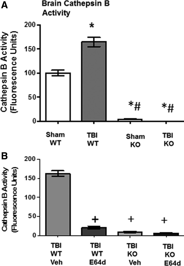
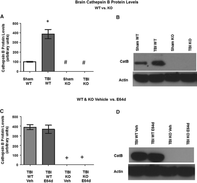
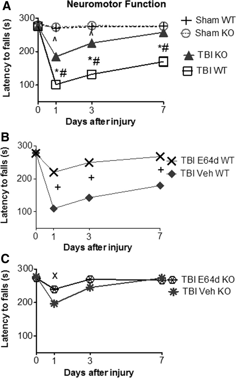
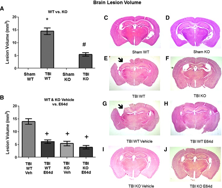
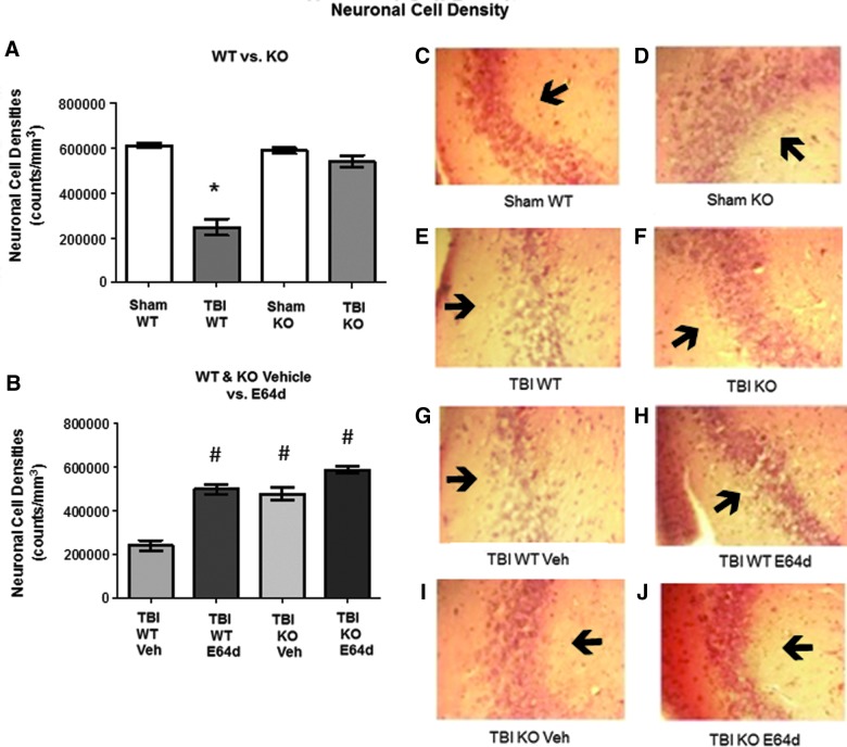
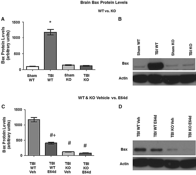
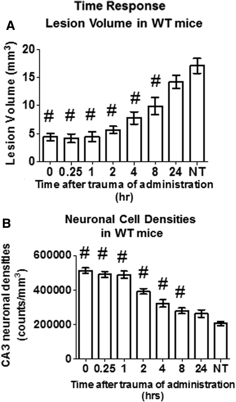
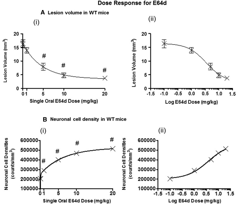
There are currently no effective therapeutic agents for traumatic brain injury (TBI), but drug treatments for TBI can be developed by validation of new drug targets and demonstration that compounds directed to such targets are efficacious in TBI animal models using a clinically relevant route of drug administration. The cysteine protease, cathepsin B, has been implicated in mediating TBI, but it has not been validated by gene knockout (KO) studies. Therefore, this investigation evaluated mice with deletion of the cathepsin B gene receiving controlled cortical impact TBI trauma. Results indicated that KO of the cathepsin B gene resulted in amelioration of TBI, shown by significant improvement in motor dysfunction, reduced brain lesion volume, greater neuronal density in brain, and lack of increased proapoptotic Bax levels. Notably, oral administration of the small-molecule cysteine protease inhibitor, E64d, immediately after TBI resulted in recovery of TBI-mediated motor dysfunction and reduced the increase in cathepsin B activity induced by TBI. E64d outcomes were as effective as cathepsin B gene deletion for improving TBI. E64d treatment was effective even when administered 8 h after injury, indicating a clinically plausible time period for acute therapeutic intervention. These data demonstrate that a cysteine protease inhibitor can be orally efficacious in a TBI animal model when administered at a clinically relevant time point post-trauma, and that E64d-mediated improvement of TBI is primarily the result of inhibition of cathepsin B activity. These results validate cathepsin B as a new TBI therapeutic target.


Figures

<b>FIG. 1.</b>
FIG. 1.
TBI increases brain cathepsin B activity, which is eliminated by cathepsin B gene KO and is reduced by oral E64d administration. Brain cathepsin B activities were determined 2 h after sham operation and TBI trauma, expressed as fluorescent units per mg protein. (A) Brain cathepsin B activities for sham and TBI conditions in WT and cathepsin B KO animals are shown. Mean cathepsin B activities differ among groups, with TBI WT mice having a higher brain cathepsin B activity than all other groups (*) and sham KO and TBI KO mice having a lower brain cathepsin B activity than sham WT mice (#). (B) Brain cathepsin B activities of TBI WT and cathepsin B KO mice treated orally with either E64d (10 mg/kg) or Veh immediately after TBI trauma (5–10 min) are shown. Mean cathepsin B activities differ among groups, with E64d TBI WT, vehicle-treated TBI KO, and E64d-treated KO mice having lower brain cathepsin B activities than TBI WT vehicle-treated mice (+) (N=10 animals/group; significant differences, p<0.05, Bonferroni's multiple comparison test). TBI, traumatic brain injury; KO, knockout; WT, wild type; Veh, vehicle.
<b>FIG. 2.</b>
FIG. 2.
TBI increases brain cathepsin B protein levels, which is eliminated by cathepsin B gene KO and is not affected by E64d treatment. Brain cathepsin B protein levels were determined 24 h after sham operation or TBI trauma. (A) Quantitative analysis of brain cathepsin B protein levels of sham and TBI conditions in WT and cathepsin B KO mice are shown. TBI WT mice had higher mean cathepsin B levels than all other groups (*), and sham KO and TBI KO mice had lower cathepsin B levels than sham WT mice (#). (B) An exemplary Western blot of brain cathepsin B protein levels of sham and TBI conditions in WT and KO mice is shown. (C) Quantitative comparisons of brain cathepsin B protein levels of the TBI condition in WT and KO mice treated with vehicle or E64d (10 mg/kg) immediately after trauma. TBI KO mice treated with either vehicle or E64d had lower levels than TBI WT mice treated with either vehicle or E64d (+). (D) An exemplary Western blot of brain cathepsin B levels of the TBI condition in WT and KO mice, treated with vehicle or E64d (N=10 animals/group; significant differences, p<0.05, Bonferroni's multiple comparison test). TBI, traumatic brain injury; KO, knockout; WT, wild type; Veh, vehicle.
<b>FIG. 3.</b>
FIG. 3.
TBI causes severe neuromotor dysfunction, which is improved by cathepsin B gene KO and by oral E64d treatment. Neuromotor dysfunction was assessed at different time points during the week after TBI trauma using the rotarod assay by measuring latency to fall time, with a shorter time reflecting a greater dysfunction. (A) Latency to fall times for sham and TBI conditions in WT and cathepsin B KO mice are shown. At day 0 (pretrauma), there was no difference in performance among groups. At all days post-trauma, there was no difference between sham WT and sham KO mice. In contrast, there were significant differences in latency to fall times among groups on days 1, 3, and 7 post-trauma. TBI WT mice had shorter times than for sham WT mice (*) on days 1, 3, and 7 post-trauma, showing that TBI caused significant impairment and a failure to recover within the measured time period. TBI KO mice had shorter times than sham KO mice on days 1 and 3 post-trauma (^), but by day 7, TBI KO mice regained the function of the sham animals and thus had recovered within the measured time period. Importantly, cathepsin B KO mice had longer times than TBI WT mice on days 1, 3, and 7 post-trauma (#), showing that cathepsin B gene KO reduced the severity of motor dysfunction on all days post-trauma. (B) Latency to fall times for TBI WT mice treated with E64 (10 mg/kg) or Veh immediately after trauma are shown. E64d treatment of TBI WT mice resulted in significantly improved motor performances on all days post-trauma, compared to Veh-treated animals (+). TBI WT Veh-treated and TBI WT mice (from panel A) had equivalent performances. (C) Latency to fall times for TBI KO mice treated with E64d (10 mg/kg) or Veh immediately after trauma are shown. E64d treatment of TBI KO mice resulted in longer latency time on day 1, relative to Veh treatment (×), but on days 3 and 7, there was no significant difference in times for E64d- and Veh-treated animals. No significant difference was observed in mean performances on all days between TBI KO Veh-treated mice and TBI KO mice (from panel A) or between the TBI KO E64d-treated and TBI WT E64d-treated mice (from panel B) (N=10 animals/group; significant differences, p<0.05, Bonferroni's multiple comparison test). TBI, traumatic brain injury; KO, knockout; WT, wild type; Veh, vehicle.
<b>FIG. 4.</b>
FIG. 4.
Brain lesion caused by TBI is reduced by cathepsin B gene KO and by oral E64d treatment. Animals tested in the rotarod assay were sacrificed at day 7 post-TBI and their were brains analyzed by quantitative histology to determine brain lesion volume at the impact site. (A) Quantitative brain lesion volumes for sham and TBI conditions in WT and KO mice are shown. TBI WT mice had a significantly larger lesion volume than sham WT, sham KO, and TBI KO mice (*), which had a larger volume than either sham groups (#). (B) Quantitative brain lesion volumes for the TBI condition in WT and KO mice treated with vehicle or E64d are shown. E64d-treated TBI WT, Veh-treated TBI KO, and E64d-treated TBI KO mice had significantly lower lesion volumes than Veh-treated TBI WT mice (+). There was no difference in volumes between Veh-treated or untreated TBI WT mice or between E64d- and Veh-treated TBI KO mice and untreated TBI KO mice (A). (C–J) Exemplary brain micrographs from each group are shown. Note the absence of brain tissue in the upper-left quadrant of (E) and (G), which are from TBI WT and TBI WT vehicle-treated animals, respectively (arrow; full picture width, 10 mm; N=10 animals/group; significant differences, p<0.05, Bonferroni's multiple comparison test). TBI, traumatic brain injury; KO, knockout; WT, wild type; Veh, vehicle. Color image is available online at www.liebertpub.com/neu
<b>FIG. 5.</b>
FIG. 5.
TBI-mediated reduction of hippocampal neuronal densities in WT mice is restored by cathepsin B gene KO and by oral E64d treatment. Brains of animals analyzed in the rotarod assay were also evaluated at day 7 post-TBI for neuronal cell densities in the CA3 region of the hippocampus by quantitative histological methods. (A) Quantitative neuronal cell densities for sham and TBI conditions in WT and KO mice are shown. TBI WT mice had significantly less neuronal cell density than all other groups (*). (B) Quantitative neuronal cell densities for the TBI condition of WT and KO mice treated with E64d or Veh treatment are shown. E64d-treated WT and KO mice and vehicle-treated KO mice had increased neuronal cell densities, compared to Veh-treated TBI WT mice (#). E64d- and vehicle-treated TBI KO mice and E64d-treated WT mice had the same densities as the untreated TBI KO and sham KO and WT mice (A). (C–J) Exemplary micrographs of the hippocampal region from each group are shown. Dark spots within micrographs are neurons, which are aggregated together to form the dark band of the CA3 layer (arrow). Note that the density of the layer is much less in the TBI WT micrograph than in the TBI KO micrograph (E and F) and is much less in the TBI WT Veh vs. TBI WT E64d micrographs (I and H) (full picture width, 0.1 mm; N=10 animals/group; significant differences, p<0.05, Bonferroni's multiple comparison test). TBI, traumatic brain injury; KO, knockout; WT, wild type; Veh, vehicle. Color image is available online at www.liebertpub.com/neu
<b>FIG. 6.</b>
FIG. 6.
TBI-induced elevation of Bax levels in brains is reduced by cathepsin B KO and E64d treatment. Brains of animals used to measure cathepsin B protein levels (Fig. 2) were also used to measure proapoptotic Bax protein levels by quantitative densitometry of western blots. (A) Brain Bax levels of sham and TBI conditions in WT and KO mice are shown. Mean levels were different among the groups, with TBI WT mice having increased Bax levels relative to all other groups (*). (B) An exemplary western blot of brain Bax protein levels from sham and TBI conditions in WT and KO mice is shown. (C) Brain Bax levels are shown for the TBI condition in WT and KO mice treated with E64d (10 mg/kg) or Veh immediately after trauma. Mean levels in TBI WT Veh-treated mice were higher level than all other groups (#), indicating that E64d and cathepsin B KO reduced the TBI-induced increase in Bax levels. E64d TBI WT mice had higher levels than either treatment of the KO mice (+). (E64d- or Veh-treated TBI KO mice had the same densities as untreated sham and TBI KO mice and sham WT mice [A].) (D) An exemplary western blot is shown of the TBI condition in WT and KO mice, with E64d or Veh treatment (N=10 animals/group; significant differences, p<0.05, Bonferroni's multiple comparison test). TBI, traumatic brain injury; KO, knockout; WT, wild type; Veh, vehicle.
<b>FIG. 7.</b>
FIG. 7.
Effectiveness of a single oral E64d dose is inversely proportional to the time of administration after TBI trauma and is effective up to 8 h post-trauma at reducing brain lesion volume and increasing brain neuronal cell density. Effects of administering a single oral E64d dose (10 mg/kg) to WT mice at various times after trauma on brain lesion volume and CA3 hippocampal neuronal cell density are shown. Experimental animals and those that received no treatment (NT) were sacrificed 7 days post-trauma and compared. (A) Brain lesion volumes for groups receiving E64d at increasing times after trauma and for the NT group are shown. Lesion volume was significantly reduced for all groups receiving E64d up to 8 h post-trauma, relative to that of the NT group (#). (B) Neuronal cell densities for groups receiving E64d at increasing times after trauma and for the NT group are shown. Density was significantly increased for all groups receiving E64d up to 8 h post-trauma, relative to that of the NT group (#) (N=10 animals/group; significant differences, p<0.05, Dunnett's multiple comparison test). WT, wild type.
<b>FIG. 8.</b>
FIG. 8.
Oral E64d dose-response effects on brain lesion volume and neuronal cell density. Effects of a single oral E64d dose given to WT mice 0.5 h after trauma on brain lesion volume and CA3 hippocampal neuronal cell density were determined. (A) (i) Brain lesion volume versus the E64d dose is plotted on a linear-linear scale and fitted to a decreasing biphasic exponential curve. A steep reduction in brain lesion volume occurred for 0- to 5-mg/kg doses, followed by a gradual reduction for 5- to 20-mg/kg doses. Doses at or above 5 mg/kg resulted in significant reductions in brain lesion volumes, relative to no dose (#). (A) (ii) Brain lesion volume versus the E64d dose is plotted on a linear-logarithmic scale and fitted to a decreasing sigmoidal curve. (B) (i) Neuronal density versus E64d dose is plotted on a linear-linear scale and fitted to an increasing biphasic exponential scale. A steep increase in neuronal cell density occurred for 0- to 5-mg/kg doses, followed by a gradual reduction for 5- to 20-mg/kg doses is shown. Doses at or above 1 mg/kg resulted in significant increase in neuronal cell densities, relative to no dose (#). (B) (ii) Neuronal cell density versus the E64d dose is plotted on a linear-logarithmic scale and fitted to an increasing sigmoidal curve (N=10 animals/group; significant differences, p<0.05, Dunnett's multiple comparison test). WT, wild type.

References

    1. Menon D.K., Schwab K., Wright D.W., and Maas A.I. (2010). Position statement: definition of traumatic brain injury. Arch. Phys. Med. Rehabil. 91, 1637–1640 - PubMed
    1. Faul M., Xu L.X., Wald M., and Coronado V. (2010). Traumatic brain injury in the United States: emergency department visits, hospitalizations, and deaths. Centers for Disease Control and Prevention, National Center for Intjury Prevention and Control: Atlanta, GA
    1. Rutland-Brown W., Langlois J.A., Thomas K.E., and Xi Y.L. (2006). Incidence of traumatic brain injury in the United States, 2003. J. Head Trauma Rehabil. 21, 544–548 - PubMed
    1. Warden D. (2006). Military TBI during the Iraq and Afghanistan wars. J. Head Trauma Rehabil. 21, 398–402 - PubMed
    1. Park E., Bell J.D., and Baker A.J. (2008). Traumatic brain injury: can the consequences be stopped? CMAJ 178, 1163–1170 - PMC - PubMed

Publication types