. 2013 Dec;15(12):1664-72.
doi: 10.1093/neuonc/not112. Epub 2013 Sep 30.

Invasion patterns in brain metastases of solid cancers

Affiliations

Invasion patterns in brain metastases of solid cancers

Anna S Berghoff et al. Neuro Oncol. 2013 Dec.

Abstract

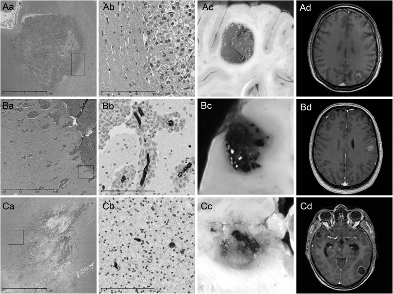
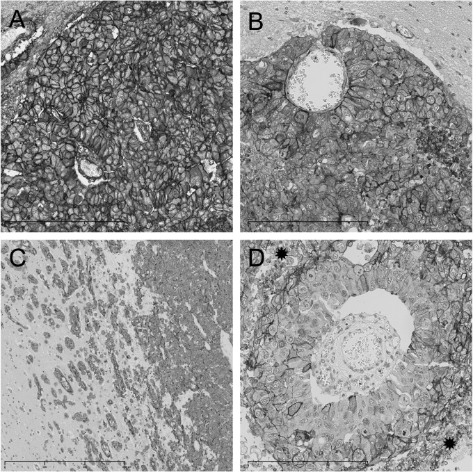
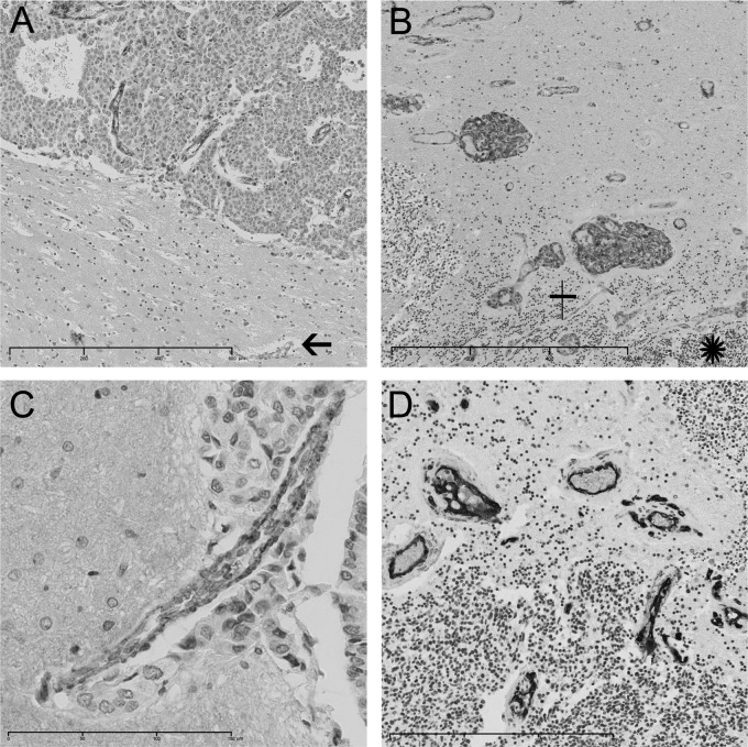
Background: Brain metastases are generally considered to be well demarcated from the surrounding brain parenchyma, although infiltrative growth patterns have been observed. We systemically investigated infiltration patterns and expression of adhesion molecules in a large and well-defined series of autopsy cases of brain metastases.

Methods: Ninety-seven autopsy specimens from 57 brain metastasis patients (primary tumor: 27 lung cancer, 6 breast cancer, 8 melanoma, 2 colorectal cancer, 1 kidney cancer, and 13 other) were evaluated for patterns of invasion into surrounding brain parenchyma. Expression of integrins αv; cytoplasmic β3, αvβ3, αvβ5, αvβ6, and αvβ8; and of E and N cadherin were evaluated using immunohistochemistry.

Results: Three main invasion patterns were seen: well-demarcated growth (29/57, 51%), vascular co-option (10/57, 18%), and diffuse infiltration (18/57, 32%). There was no statistically significant association of invasion pattern with primary tumor type, although vascular co-option was most common in melanoma brain metastases (4/10). Invasion patterns of different brain metastases of the same patient were highly concordant (P < .001, chi-square test). Distance of infiltration from the main tumor mass ranged from 12.5 µm to 450 µm (median 56.2 µm) and was not significantly different between the vascular co-option and the diffuse infiltration groups. Levels of αvβ6 were significantly higher in the well-demarcated group than in the vascular co-option and the diffuse infiltration groups (P = .033, Kruskal-Wallis test). Expression of αvβ5 in tumor cells was higher in brain metastasis lesions previously treated with stereotactic radiosurgery (P = .034, chi-square test).

Conclusions: Distinct invasion patterns of brain metastases into the brain parenchyma are not specific for primary tumor types, seem to be influenced by expression of αv integrin complexes, and may help to guide clinical decision-making.

Keywords: adhesion molecules; brain metastases; cadherin; integrin; invasion.


Figures

Fig. 1.
Fig. 1.
Distinct invasion patterns in human brain metastases. Top row: example of well-demarcated invasion (patient 5, NSCLC, Supplemental Table S1): (Aa) H&E slide (magnification ×1.25); (Ab) invasion front of well-demarcated brain metastasis (magnification × 400); (Ac) macroscopic sample of well-demarcated brain metastasis; (Ad) MRI contrast-enhanced T1-weighted sequence sample of well-demarcated brain metastasis. Middle row: example of vascular co-option (patient 13, melanoma, Supplemental Table S1): (Ba) H&E slide (magnification × 1.25); (Bb) CD34 immunohistochemistry of vascular co-option (magnification × 400); (Bc) macroscopic sample of brain metastasis growing via vascular co-option; (Bd) MRI contrast-enhanced T1-weighted sequence sample of brain metastasis growing via vascular co-option. Bottom row: example of diffuse invasion (patient 2, SCLC, Supplemental Table S1): (Ca) H&E of brain metastasis (magnification × 1.25); (Cb) CD18 cytokeratin staining showing diffusely infiltrating tumor cells; (Cc) macroscopic sample of diffuse infiltrating brain metastasis; (Cd) MRI contrast-enhanced T1-weighted sequence sample of diffuse infiltrating brain metastasis. Color figures available from authors upon request.
Fig. 2.
Fig. 2.
Integrin expression patterns: (A) αv subunit expression (patient 5, NSCLC, Supplemental Table S1); (B) αvβ5 expression with accentuation around a vessel (patient 5, NSCLC, Supplemental Table S1); (C) αvβ5 expression in a brain metastasis growing via vascular co-option (patient 13, melanoma, Supplemental Table S1); (D) αvβ6 expression with accentuation around necrosis, the stars at upper left and lower right mark the necrosis (patient 5, NSCLC, Supplemental Table S1). Color figures available from authors upon request.
Fig. 3.
Fig. 3.
Integrin expression in vascular structures: (A) αvβ3 expression on tumor vessel, arrow marks a vessel in the peritumoral area without αvβ3 expression (patient 5, NSCLC, Supplemental Table S1); (B) αvβ3 expression in glomeruloid-like vessels of the peritumoral area, the star at lower right marks the tumor tissue, the cross marks the surrounding cerebellum (patient 8, SCLC, Supplemental Table S1); (C) αvβ3 expression in a vessel with vascular co-option (patient 13, melanoma, Supplemental Table S1); (D) CD34 immunohistochemistry of the case shown in (B), illustrating the peritumoral sprouting vessels (patient 8, SCLC, Supplemental Table S1). Color figures available from authors upon request.

References

    1. Gavrilovic IT, Posner JB. Brain metastases: epidemiology and pathophysiology. J Neurooncol. 2005;75(1):5–14. - PubMed
    1. Gaspar LE, Mehta MP, Patchell RA, et al. The role of whole brain radiation therapy in the management of newly diagnosed brain metastases: a systematic review and evidence-based clinical practice guideline. J Neurooncol. 2010;96(1):17–32. - PMC - PubMed
    1. Kalkanis SN, Kondziolka D, Gaspar LE, et al. The role of surgical resection in the management of newly diagnosed brain metastases: a systematic review and evidence-based clinical practice guideline. J Neurooncol. 2010;96(1):33–43. - PMC - PubMed
    1. Linskey ME, Andrews DW, Asher AL, et al. The role of stereotactic radiosurgery in the management of patients with newly diagnosed brain metastases: a systematic review and evidence-based clinical practice guideline. J Neurooncol. 2010;96(1):45–68. - PMC - PubMed
    1. Wesseling P, von Deimling A, Aldape K. Metastatic Tumours of the CNS. 4th ed. Lyons, France: IARC Press; 2007.

Publication types

MeSH terms