

Isoflurane on brain inflammation
- PMID: 24084689
- PMCID: PMC3919516
- DOI: 10.1016/j.nbd.2013.09.016
Isoflurane on brain inflammation
Erratum in
-
Corrigendum to "Isoflurane on brain inflammation" [Neurobiology of Disease, 62 (2014) Pages 365-371].Neurobiol Dis. 2025 Jan;204:106770. doi: 10.1016/j.nbd.2024.106770. Epub 2024 Dec 17. Neurobiol Dis. 2025. PMID: 39694721 No abstract available.
Abstract
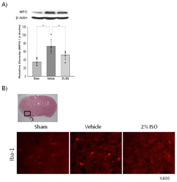
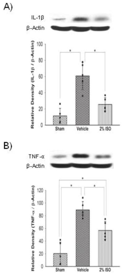
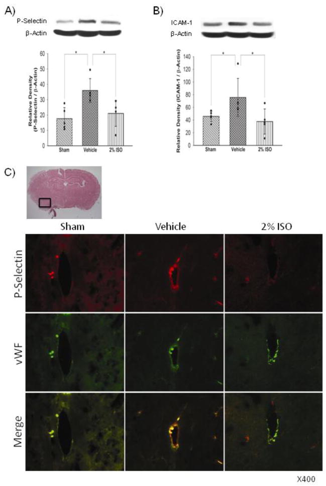
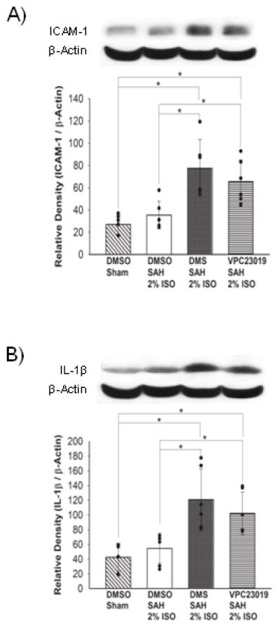
Brain inflammation may play an important role in the pathophysiology of early brain injury after subarachnoid hemorrhage (SAH). Our aim was to demonstrate brain inflammation development and to determine whether isoflurane, a clinically available volatile anesthetic agent, prevents brain inflammation after SAH. This study used 162 8-week-old male CD-1 mice. We induced SAH with endovascular perforation in mice and randomly assigned animals to sham-operated (n=21), SAH+vehicle-air (n=35) and SAH+2% isoflurane (n=31). In addition to the evaluation of brain injury (neurological scores, brain edema and Evans blue dye extravasation), brain inflammation was evaluated by means of expression changes in markers of inflammatory cells (ionized calcium binding adaptor molecule-1, myeloperoxidase), cytokines (tumor necrosis factor [TNF]-α, interleukin-1β), adhesion molecules (intercellular adhesion molecule [ICAM]-1, P-selectin), inducers of inflammation (cyclooxygenase-2, phosphorylated c-Jun N-terminal kinase [p-JNK]) and endothelial cell activation (von Willebrand factor) at 24h post-SAH. Sphingosine kinase inhibitor (N, N-dimethylsphingosine [DMS]) and sphingosine-1-phosphate receptor-1/3 antagonist (VPC23019) were used to block isoflurane's effects (n=22, each). SAH caused early brain injury, which was associated with inflammation so that all evaluated markers of inflammation were increased. Isoflurane significantly inhibited both brain injury (P<0.001, respectively) and inflammation (myeloperoxidase, P=0.022; interleukin-1β, P=0.002; TNF-α, P=0.015; P-selectin, P=0.010; ICAM-1, P=0.016; p-JNK, P<0.001; cyclooxygenase-2, P=0.003, respectively). This beneficial effect of isoflurane was abolished with DMS and VPC23019. Isoflurane may suppress post-SAH brain inflammation possibly via the sphingosine-related pathway.
Keywords: BWC; Brain water content; COX-2; Cyclooxygenase-2; DMS; EBI; Early brain injury; ICAM-1; IL-1β; Inflammation; Intercellular adhesion molecule-1; Interleukin-1beta; Ionized calcium binding adaptor molecule-1; Isoflurane; MPO; Myeloperoxidase; N, N-dimethylsphingosine; Phosphorylated c-Jun N-terminal kinase; S1P; S1P1/3; SAH; SphK; Sphingosine 1-phosphate; Sphingosine kinase; Sphingosine-1-phosphate receptor-1/3; Subarachnoid hemorrhage; TNF-α; Tumor necrosis factor-alpha; iba-1; p-JNK; vWF; von Willebrand factor.
© 2013.
Conflict of interest statement
The authors report no conflicts of interest.
Figures

References
-
- Aggarwal BB, Shishodia S, Ashikawa K, Bharti AC. The role of TNF and its family members in inflammation and cancer: lessons from gene deletion. Curr Drug Targets Inflamm Allergy. 2002;1:327–341. - PubMed
-
- Antkowiak B. How do general anaesthetics work? Naturwissenschaften. 2001;88:201–213. - PubMed
-
- Balduini W, Carloni S, Mazzoni E, Cimino M. New therapeutic strategies in perinatal stroke. Curr Drug Targets CNS Neurol Disord. 2004;3:315–323. - PubMed
Publication types
MeSH terms
Substances
Grants and funding
LinkOut - more resources
Full Text Sources
Other Literature Sources
Medical
Research Materials
Miscellaneous
