. 2013 Nov;123(11):4695-705.
doi: 10.1172/JCI71543.

Divergence of IL-1, IL-18, and cell death in NLRP3 inflammasomopathies

Divergence of IL-1, IL-18, and cell death in NLRP3 inflammasomopathies

Susannah D Brydges et al. J Clin Invest. 2013 Nov.

Abstract

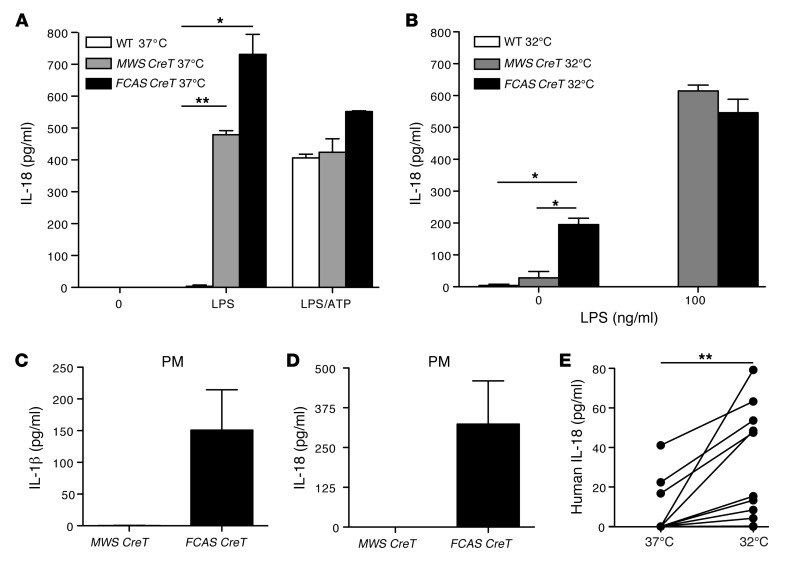
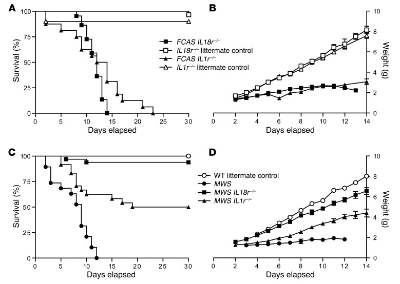
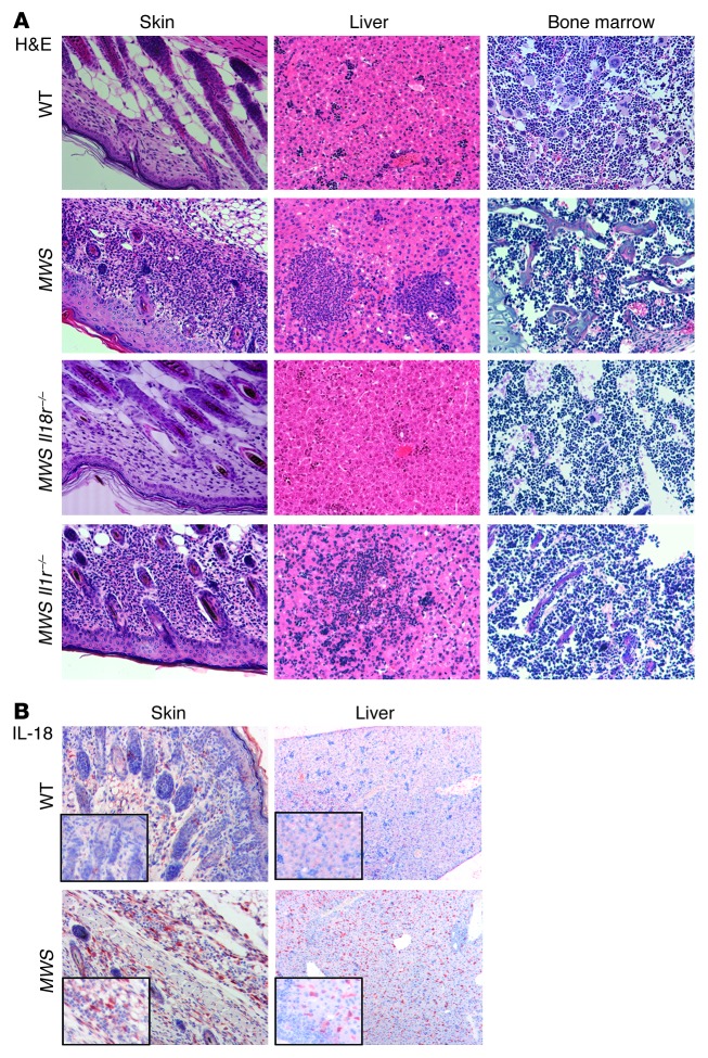
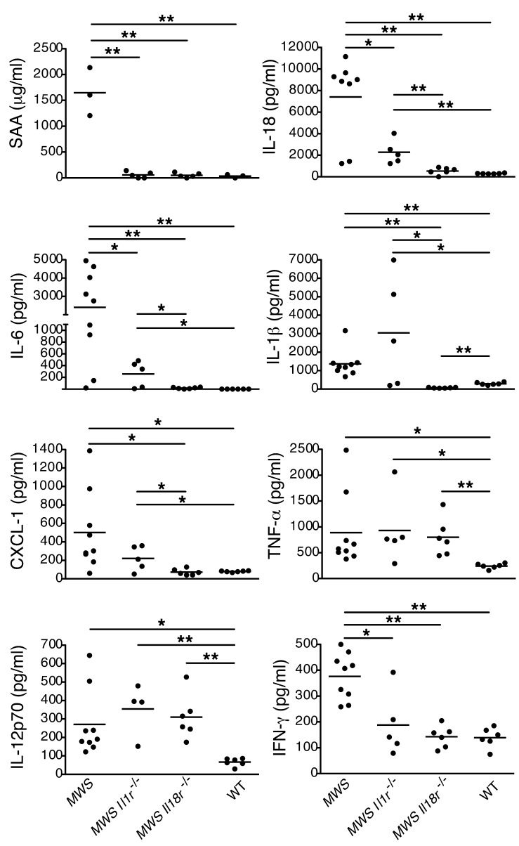
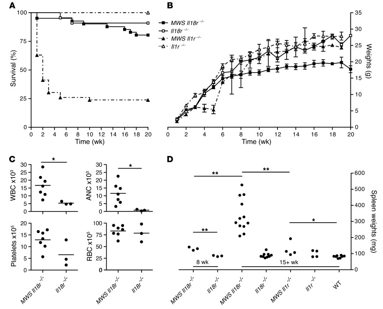
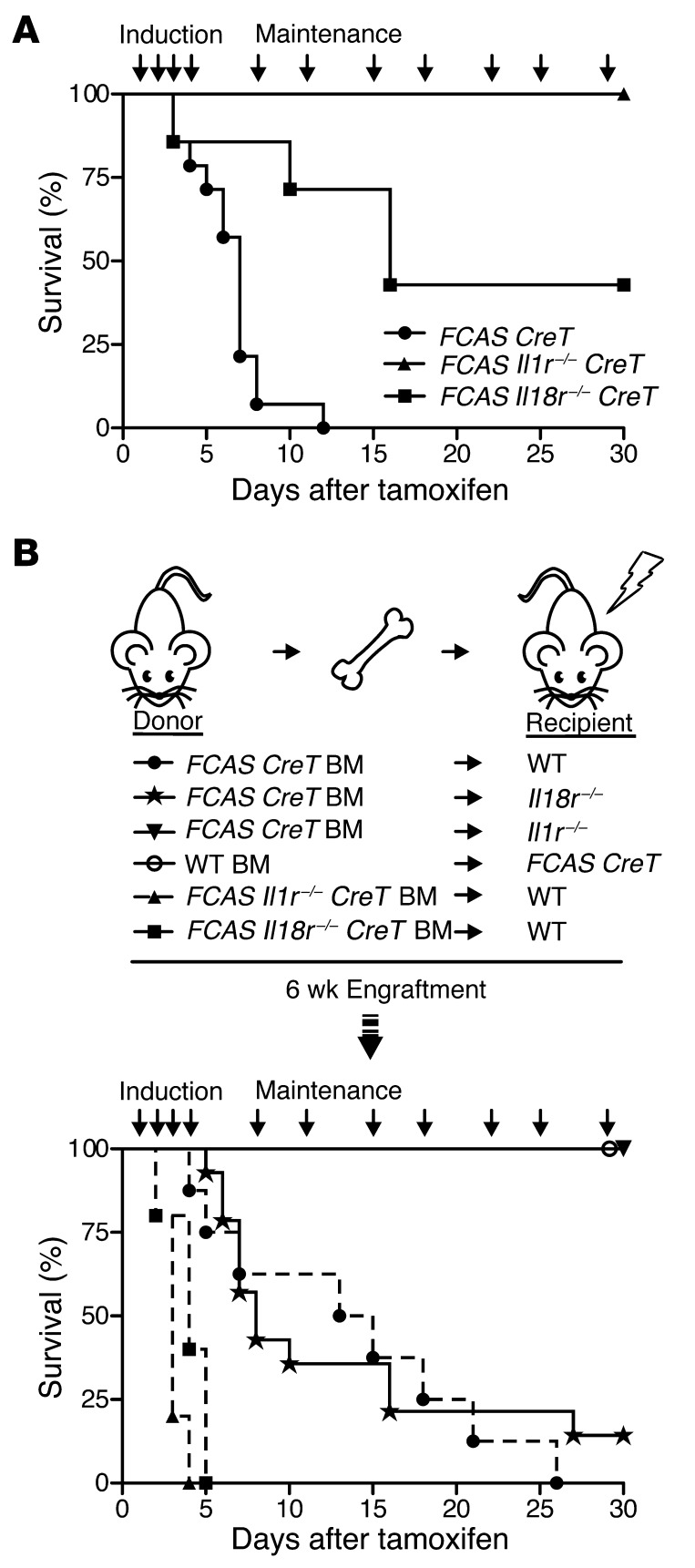
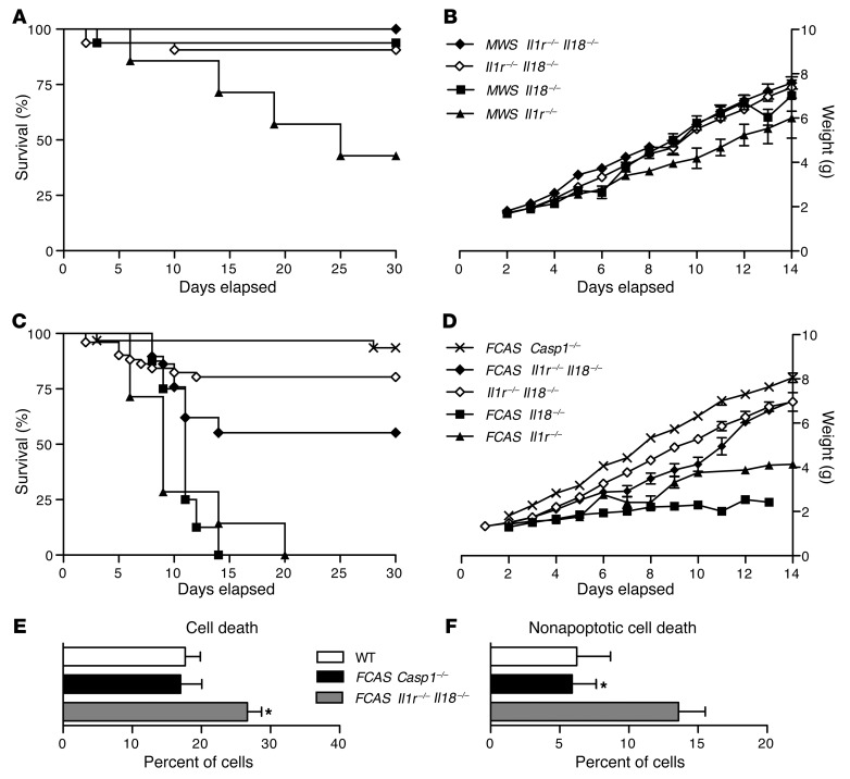
The inflammasome is a cytoplasmic multiprotein complex that promotes proinflammatory cytokine maturation in response to host- and pathogen-derived signals. Missense mutations in cryopyrin (NLRP3) result in a hyperactive inflammasome that drives overproduction of the proinflammatory cytokines IL-1β and IL-18, leading to the cryopyrin-associated periodic syndromes (CAPS) disease spectrum. Mouse lines harboring CAPS-associated mutations in Nlrp3 have elevated levels of IL-1β and IL-18 and closely mimic human disease. To examine the role of inflammasome-driven IL-18 in murine CAPS, we bred Nlrp3 mutations onto an Il18r-null background. Deletion of Il18r resulted in partial phenotypic rescue that abolished skin and visceral disease in young mice and normalized serum cytokines to a greater extent than breeding to Il1r-null mice. Significant systemic inflammation developed in aging Nlrp3 mutant Il18r-null mice, indicating that IL-1 and IL-18 drive pathology at different stages of the disease process. Ongoing inflammation in double-cytokine knockout CAPS mice implicated a role for caspase-1-mediated pyroptosis and confirmed that CAPS is inflammasome dependent. Our results have important implications for patients with CAPS and residual disease, emphasizing the need to explore other NLRP3-mediated pathways and the potential for inflammasome-targeted therapy.


Figures

Figure 1
Figure 1. Myeloid cells expressing mutant NLRP3 proteins secrete IL-18.
Tamoxifen-treated MWS CreT, FCAS CreT, and littermate WT BMDCs were incubated with pure LPS, with and without ATP at (A) 37°C or (B) 32°C. IL-18 in the supernatants was measured by ELISA (n = 4, mean and SEM). Tamoxifen-treated MWS CreT and FCAS CreT peritoneal macrophages (PM) were incubated at 32°C, and (C) IL-1β and (D) IL-18 were measured in the supernatants by ELISA (n = 2, mean and SEM). (E) Adherent monocytes from patients with FCAS were incubated at 37°C or 32°C, and IL-18 was measured in the culture supernatants by ELISA (n = 10). *P < 0.05, **P < 0.005, by Student’s t test.
Figure 2
Figure 2. IL-18R deficiency ameliorates inflammation in murine CAPS.
(A) Survival and (B) growth of FCAS Il18r–/–, Il18r–/–, FCAS ll1r–/–, and Il1r–/– mice (n = 16–30 for survival and 1–29 for growth). (C) Survival and (D) growth of MWS, MWS Il18r–/–, MWS Il1r–/–, and WT littermates (n = 4–33 for survival and 1–27 for growth). Statistical significance was tested by log-rank (Mantel-Cox) comparison. Error bars shown for mean daily weights on growth curves are SEM.
Figure 3
Figure 3. IL-18R signaling drives skin and liver pathology in murine CAPS.
(A) H&E-stained tissue sections from skin, liver, and bone marrow from 9-day-old littermate WT, MWS, MWS Il18r–/–, and MWS Il1r–/– mice (original magnification, ×20). (B) Immunohistochemistry on skin and liver from WT and MWS pups for IL-18 followed by hematoxylin staining (original magnification, ×10; ×40 [insets]). Sections are representative of at least 3 mice per strain.
Figure 4
Figure 4. Disruption of IL-18 signaling normalizes key CAPS serum cytokine and SAA levels.
Multiplex cytokine and ELISA analysis of serum obtained at days 6–8 from WT, MWS, MWS Il1r–/–, and MWS Il18r–/– pups (n ≥ 5 mice); each graph point represents 1 mouse, with mean identified by horizontal bar. *P < 0.05, **P < 0.005 by Student’s t test.
Figure 5
Figure 5. Aging mutant CAPS mice on IL-18R knockout background develop systemic inflammation.
(A) Extended survival and (B) growth of MWS Il18r–/–, Il18r–/–, MWS Il1r–/–, and Il1r–/– mice (n = 7–46 for survival and 1–38 for growth, mean ± SEM). Statistical significance was tested by log-rank (Mantel-Cox) comparison. (C) Complete peripheral blood counts for MWS Il1r–/– and Il18r–/– mice at 16 weeks of age (n ≥ 3 mice; each graph point represents 1 mouse, with mean identified by horizontal bar). (D) Spleens from MWS Il18r–/–, Il18r–/–, MWS Il1r–/–, Il11r–/–, and WT littermates were weighed at 8 or 15 weeks (n = 3–12 mice; each graph point represents 1 mouse, with mean identified by horizontal bar). *P < 0.05, **P < 0.005, by Student’s t test. WBC, white blood cells; RBC, red blood cells; ANC, absolute neutrophil count.
Figure 6
Figure 6. Inducible and chimeric mutant CAPS models confirm age-related cytokine dependence.
(A) Survival of FCAS CreT, FCAS Il18r–/–, or FCAS Il1r–/– mice, displayed as days after initiation of tamoxifen treatment (n = 4–14 mice). (B) Schematic of bone marrow transplants performed. Survival of transplanted mice, displayed as days after initiation of tamoxifen treatment (n = 5–14 mice). P values by log-rank (Mantel-Cox) comparison.
Figure 7
Figure 7. Caspase-1 induces inflammation in CAPS via cytokine mediators and pyroptosis.
(A) Survival and (B) growth of MWS ll1r–/–Il18–/–, Il1r–/–Il18–/–, MWS Il18–/–, and MWS Il1r–/– mice (n = 7–32 for survival and 1–29 for growth). (C) Survival and (D) growth of FCAS Casp1–/–, FCAS Il1r–/–Il18–/–, Il1r–/–Il18–/–, FCAS Il18–/–, and FCAS Il1r–/– mice (n = 7–51 for survival and 1–40 for growth). Weights are shown as mean ± SEM. Bone marrow cells were isolated from 16-week-old FCAS Casp1–/–, FCAS Il1r–/–Il18–/–, and WT mice and evaluated for (E) membrane asymmetry and (F) dye exclusion by flow cytometry. Data are representative of 3 independent experiments (mean ± SEM shown). *P < 0.05, by Student’s t test.

References

    1. Aksentijevich I, et al. The clinical continuum of cryopyrinopathies: novel CIAS1 mutations in North American patients and a new cryopyrin model. Arthritis Rheum. 2007;56(4):1273–1285. doi: 10.1002/art.22491. - DOI - PMC - PubMed
    1. Hoffman HM, Mueller JL, Broide DH, Wanderer AA, Kolodner RD. Mutation of a new gene encoding a putative pyrin-like protein causes familial cold autoinflammatory syndrome and Muckle-Wells syndrome. Nat Genet. 2001;29(3):301–305. doi: 10.1038/ng756. - DOI - PMC - PubMed
    1. Martinon F, Burns K, Tschopp J. The inflammasome: a molecular platform triggering activation of inflammatory caspases and processing of proIL-beta. Mol Cell. 2002;10(2):417–426. doi: 10.1016/S1097-2765(02)00599-3. - DOI - PubMed
    1. Martinon F, Petrilli V, Mayor A, Tardivel A, Tschopp J. Gout-associated uric acid crystals activate the NALP3 inflammasome. Nature. 2006;440(7081):237–241. doi: 10.1038/nature04516. - DOI - PubMed
    1. Cassel SL, et al. The Nalp3 inflammasome is essential for the development of silicosis. Proc Natl Acad Sci U S A. 2008;105(26):9035–9040. doi: 10.1073/pnas.0803933105. - DOI - PMC - PubMed

Publication types

MeSH terms