Review
. 2013 Nov;44(11):3275-85.
doi: 10.1161/STROKEAHA.113.000736. Epub 2013 Oct 1.

Brain regulation of thrombosis and hemostasis: from theory to practice

Affiliations
Review

Brain regulation of thrombosis and hemostasis: from theory to practice

Mark J Fisher. Stroke. 2013 Nov.
No abstract available

Keywords: blood-brain barrier; hemorrhage; hemostasis; thrombosis.


Figures

Figure 1
Figure 1
The complexities of the coagulation pathways are illustrated here. The effects of tissue factor (TF)-mediated activation of factor VII (FVIIa), representing extrinsic pathway activation, are amplified by the intrinsic pathway; this results in thrombin activation and generation of soluble fibrin monomer (SFM) and fibrin clot. This procoagulant pathway is negatively regulated by three anticoagulant pathways, two of which are derived from endothelial cells (EC): tissue factor pathway inhibitor (TFPI), which forms a quaternary complex with TF, FVIIa, and Factor Xa (FXa); and the thrombomodulin (TM)-protein C (PC) pathway in which the TM-thrombin complex activates PC, a process which is amplified by the endothelial protein C receptor (EPCR), with the resulting activated protein C (aPC) and its cofactor protein S (PS) then capable of inactivating factor Va (FVa) and factor VIIIa (FVIIIa). The third major anticoagulant pathway consists of circulating antithrombin III (AT), which can inhibit thrombin, factor Xa (FXa), and other serine proteases; actions of AT are vastly amplified by its binding with endothelial-derived heparan sulfate proteoglycans (not shown). FXa is also inhibited by protein Z (PZ), while heparin cofactor (HC II) is another thrombin inhibitor. Fibrin dissolution is produced by the fibrinolytic pathway, with plasmin-induced clot lysis producing fibrin degradation products (FDP). Plasmin is derived from tissue-type plasminogen activator (tPA) and/or urokinase-type plasminogen activator (uPA) actions on plasminogen. Both tPA and uPA are inhibited by plasminogen activator inhibitor-1 (PAI-1), while tPA effects are amplified by annexin II (AnxII). Other negative regulators of fibrinolysis include α2-antiplasmin (α2-AP) and thrombin-activatable fibrinolysis inhibitor (TAFI). Thrombogenic surfaces for the factor Va (FVa):factor Xa (FXa) (prothrombinase) complex, the factor VIIIa (FVIIIa):factor IXa (FIXa) (intrinsic tenase) complex, and for factor XIa (FXIa) are provided by platelets (Plt). Factor XIIa (FXIIa) may participate in initiation and/or propagation of clot, while factor XIIIa stabilizes thrombus by cross-linking fibrin monomers. (Reprinted with permission from Mackman N: Tissue specific hemostasis in mice. Arteroscler Thromb Vasc Biol. 2005;25:2273–2281)
Figure 2
Figure 2
Endothelial tight junctions, with transmembrane molecules occludin and claudin interacting with actin cytoskeleton and zonula occludens proteins including ZO-1. (From Kim JH, Kim JH, Park JA, Lee SW, Kim WJ, Yu YS, Kim KW. Blood-neural barrier: intercellular communication at glio-vascular interface. J Biochem Mol Biol. 2006;39:339–345, copyright BMB Reports, http://www.bmbReports.org/)
Figure 3
Figure 3
A model illustrating elements of brain-specific hemostasis regulation. A pericyte, opposite capillary tight junction, prevents red blood cell exit while tissue-factor expressing astrocytes provide additional protection against hemorrhage.
Figure 4
Figure 4
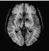
Examples of severe cerebral microbleeds, imaged using 3Tesla MRI and susceptibility-weighted imaging sequences. 4A is from a 67 year old male with hypertension, 4B is from a 51 year old male with multiple medical problems including hypertension, diabetes, end-stage renal disease, sepsis, and thrombocytopenia; a right frontal intracerebral hemorrhage was also present (not shown).
Figure 4
Figure 4
Examples of severe cerebral microbleeds, imaged using 3Tesla MRI and susceptibility-weighted imaging sequences. 4A is from a 67 year old male with hypertension, 4B is from a 51 year old male with multiple medical problems including hypertension, diabetes, end-stage renal disease, sepsis, and thrombocytopenia; a right frontal intracerebral hemorrhage was also present (not shown).
Figure 5
Figure 5
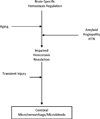
A model relating brain-specific hemostasis regulation to development of cerebral microscopic hemorrhage, cerebral microbleeds, and white matter disease. Transient injury may be inflammatory, and pathways leading to white matter disease include variants of blood-brain barrier injury that are not necessarily a direct consequence of microbleeds. (HTN: hypertension)

References

    1. Aird WC. Coagulation. Crit Care Med. 2005;33(Suppl):S485–S487. - PubMed
    1. del Zoppo GJ. Virchow’s triad: The vascular basis of cerebral injury. Rev Neurol Dis. 2008;5(Suppl 1):S12–S21. - PMC - PubMed
    1. Wolberg AS, Aleman MM, Leiderman K, Machlus KR. Procoagulant activity in hemostasis and thrombosis: Virchow’s triad revisited. Anesth Analg. 2012;114:275–285. - PMC - PubMed
    1. Mackman N. Tissue-specific hemostasis in mice. Arterioscler Thromb Vasc Biol. 2005;25:2273–2281. - PubMed
    1. Rosenberg RD, Aird WC. Vascular-bed-specific hemostasis and hypercoagulable states. N Engl J Med. 1999;340:1555–1564. - PubMed

Publication types

MeSH terms