. 2013 Nov;27(11):1934-45.
doi: 10.1210/me.2013-1099. Epub 2013 Oct 1.

Melanocortin 4 receptor becomes an ACTH receptor by coexpression of melanocortin receptor accessory protein 2

Affiliations

Melanocortin 4 receptor becomes an ACTH receptor by coexpression of melanocortin receptor accessory protein 2

Maria Josep Agulleiro et al. Mol Endocrinol. 2013 Nov.

Abstract

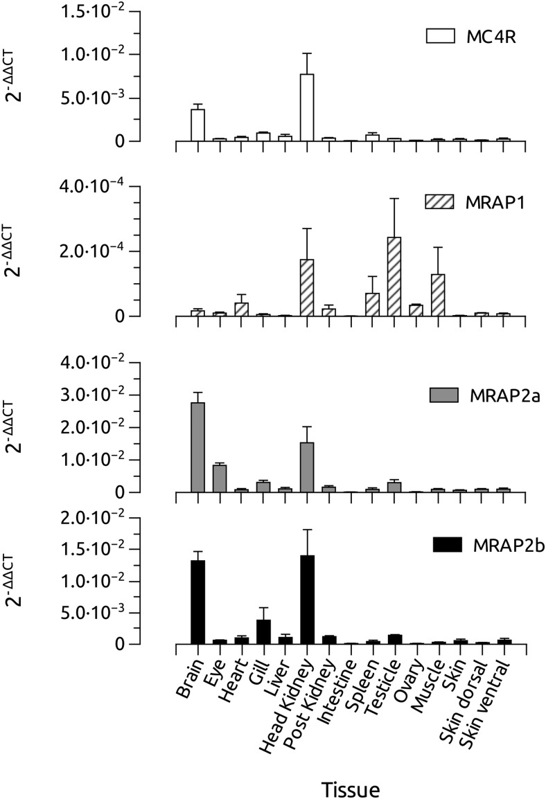
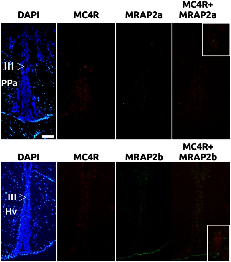
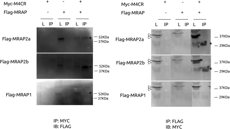
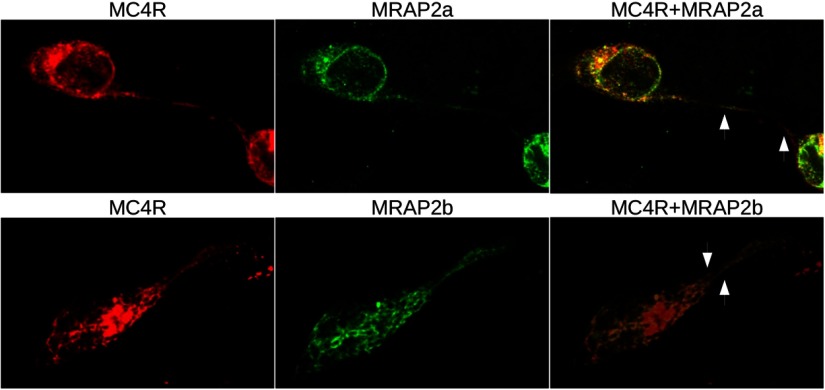
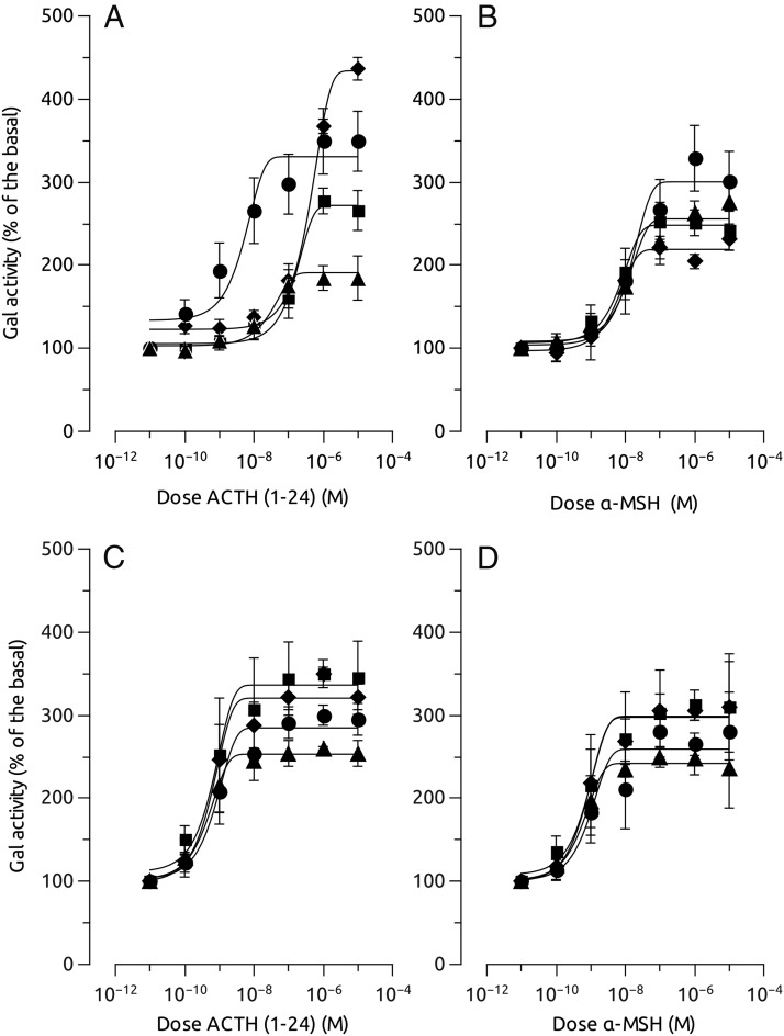
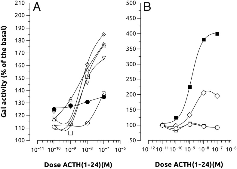
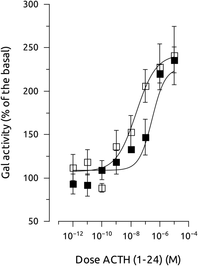
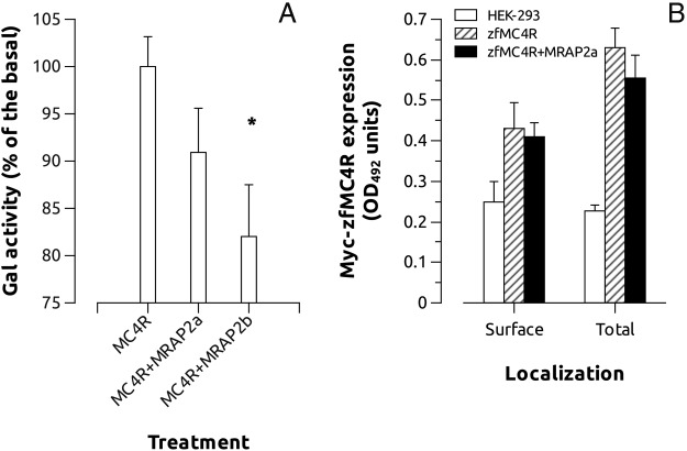
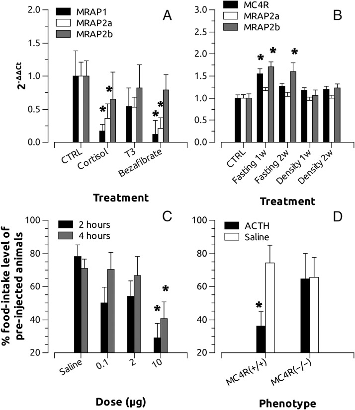
Melanocortin 2 receptor (MC2R) is the only canonical ACTH receptor. Its functional expression requires the presence of an accessory protein, known as melanocortin receptor 2 accessory protein 1 (MRAP1). The vertebrate genome exhibits a paralogue gene called MRAP2, which is duplicated in zebrafish (MRAP2a and MRAP2b), although its function remains unknown. In this paper, we demonstrate that MRAP2a enables MC4R, a canonical MSH receptor, to be activated by ACTH with a similar sensitivity to that exhibited by MC2R. Both proteins physically interact and are coexpressed in the neurons of the preoptic area, a key region in the control of the energy balance and hypophyseal secretion in fish. ACTH injections inhibit food intake in wild-type zebrafish but not in fish lacking functional MC4R. Both MRAP1 and MRAP2a are hormonally regulated, suggesting that these proteins are substrates for feed-back regulatory pathways of melanocortin signaling. Fasting has no effect on the central expression of MRAP2a but stimulates MRAP2b expression. This protein interacts and is colocalized with MC4R in the tuberal hypothalamic neurons but has no effect on the pharmacologic profile of MC4R. However, MRPA2b is able to decrease basal reporter activity in cell lines expressing MC4R. It is plausible that MRAP2b decreases the constitutive activity of the MC4R during fasting periods, driving the animal toward a positive energy balance. Our data indicate that MRAP2s control the activity of MC4R, opening up new pathways for the regulation of melanocortin signaling and, by extension, for the regulation of the energy balance and obesity.


Figures

Figure 1.
Figure 1.
Distribution of MC4R and MRAPs mRNA expression in different zebrafish tissues, as revealed by quantitative real-time PCR (qPCR). Amplifications of β-actin and 18S and EF1α mRNAs were used as internal control of the reverse transcription.
Figure 2.
Figure 2.
Double in situ hybridization of MC4R (red) and MRAP2a (green) at the level of preoptic area (upper panels) or MC4R (red) and MRAP2b (green) at the level of tuberal hypothalamus (lower panels). Samples were aslo stained with 4′6-diamidino-2-phenylindole (DAPI) for morphologic studies. III, third ventricle; HV, ventral hypothalamus; Ppa, anterior preoptic area. Insets in the right panels show magnification of MC4R and MRAP colocalization. Scale bar, 200 μm.
Figure 3.
Figure 3.
MRAP2s and MC4R interactions. HEK cells were transfected with Flag-tagged MRAPs and/or Myc-tagged MC4R. Whole-cell lysates were prepared 24 hours after transfection and used for Western blot or incubated with anti-FLAG magnetic beads or anti-MYC agarose beads for coimmunoprecipitation. Both Flag-MRAP2a and Flag-MRAP2b, but not Flag-MRAP1, interact with Myc-MC4R as seen by immunoblotting (IB) with anti-Flag and anti-Myc, respectively after immunoprecipitation (IP) with anti-Myc. Asterisks indicate positive bands in crude lysates and immunoprecipitated samples whereas white arrowheads show unspecific bands.
Figure 4.
Figure 4.
Immunofluorescence assays in live cells showing the expression of MRAPs (green) and/or zfMC4R (red). N-terminally Flag-tagged MRAPs and N-terminally Myc-tagged MC4R were transiently expressed in HEK-293 cells. Photomicrographs are taken with a ×60 objective and are from a single optical section obtained within an acquisition of z stacks (0.10 μm/slice). Arrows indicate regions of the plasma membrane where MRAPs and MC4R are potentially colocalized.
Figure 5.
Figure 5.
Pharmacologic properties of melanocortin agonist, α-MSH and hACTH (1–24) at HEK-293 transiently expressing both MC4R (upper panels) or MC5Ra (lower panels) and different MRAPs (■, MC4R; ♦, MC4R+MRAP1; •, MC4R+MRAP2a; ▴, MC4R+MRAP2b) but stably expressing a cAMP-responsive β-galactosidase reporter gene. Data were normalized to protein levels and expressed as percentage of the basal levels. A construct carrying luciferase gene under the control of a constitutive promoter was also transfected to standardize the transfection levels. Experiments were performed using quadruplicate data points and repeated at least 3 times independently. Data are mean ± SEM of the 3 independent experiments.
Figure 6.
Figure 6.
Effects of different MRAP combination on MC4R-induced galactosidase activity. A, Pharmacologic properties of hACTH(1–24) at HEK-293 transiently expressing both MC4Rs with different combinations of MRAPs (○, MC4R; □, MC4R+MRAP2a; ♢, MC4R+MRAP2a+MRAP1; ▵, MC4R+MRAP2a+MRAP2b; ▿, MC4R+MRAP2a+MRAP2b +MRAP1; •, MC4R+MRAP2b+MRAP1, and stably expressing a cAMP-responsive β-galactosidase reporter gene. Only when MRAP2a was present in the combination, the MC4R was able to respond to ACTH stimulation. B, Pharmacologic properties of hACTH(1–24) at HEK-293 transiently expressing both MC2R with different MRAPs (○, MC2R; ■, MC2R+MRAP1; □, MC2R+MRAP2a; ♢, MC2R+MRAP1+MRAP2a). Data were normalized to protein levels and expressed as percentage of the basal levels. Error bars were omitted to facilitate the graph vision. A construct carrying luciferase gene under the control of a constitutive promoter was also transfected to standardize transfection levels. Experiments were performed using quadruplicate data points and repeated 2 times independently.
Figure 7.
Figure 7.
Effects of AGRP on hACTH(1–24)-stimulated galactosidase activity in HEK-293 cells transiently expressing both MC4R and MRAP2a but stably expressing a cAMP-responsive β-galactosidase reporter gene (□, MC4R+MRAP2a; ■, MC4R+MRAP2a+AGRP). Data were normalized to protein levels and expressed as percentage of the basal levels. A construct carrying luciferase gene under the control of a constitutive promoter was also transfected to standardize transfection levels. Experiments were performed using quadruplicate data points and repeated at least 2 independent times.
Figure 8.
Figure 8.
A, Effects of MRAP2a and MRAP2b on MC4R-induced galactosidase basal activity in HEK-293 cells stably expressing MC4R but transiently expressing galactosidase gene under the control of a constitutive promoter carrying several CRE sites (see Material and Methods for more details). A construct carrying luciferase gene under the control of a constitutive promoter was also transfected to standardize transfection levels. Experiments were performed using quadruplicate data points and repeated 4 independent times. Asterisks show significant differences after Student's t test (P < .05). B, Total and cell surface detection of Myc-zfMC4R using anti-Myc antibodies. Control corresponds to nontransfected HEK-293 cells. Cells were transiently transfected with MC4R or MC4R+MRAP2a and assayed for total and extracellular c-Myc detection by whole-cell ELISA. The results represent the mean ± SEM of 3 independent experiments, each performed in triplicate.
Figure 9.
Figure 9.
A, Effects of cortisol, T3, or bezafibrate, a PPAR agonist, on MRAP expression. Animals were fed with food pellets containing 500 μg/g of the different hormones and humanely destroyed after 7 days. Total RNA of the whole fish was purified and used for cDNA synthesis. MRAP1, MRAP2a, and MRAP2b expression was analyzed with the ΔΔCt (cycle threshold) method. B, Fasting and rearing density effects on brain expression of MRAP2a and MRAP2b. Animals were fed for 14 days at 4% of the body weight and subsequently fasted for 7 and 14 days. For density experiments animals were maintained during 7 and 14 days in 1/20 of water volume when compared with the control (CTRL) group. MRAP2s expression data were treated as before. C, Effects of hACTH(1–24) on zebrafish (wild-type TU strain) food intake levels. Food intake levels were recorded during 4 consecutive days to establish a base line for each intact fish. The fifth day animals were injected ip with hACTH(1–24). After 15 minutes, food intake levels of each treated fish were recorded after 2 and 4 hours and expressed as the percentage of the base line (average of the food intake levels during the previous 4 days). D, Effects of hACTH(1–24) on zebrafish sa122 food intake levels. Food intake levels were recorded and calculated as before (see Figure 12). Asterisks show significant differences after one-way ANOVA and Tukey's method (P < .05).

References

    1. Nakanishi S, Inoue A, Kita T, et al. . Nucleotide sequence of cloned cDNA for bovine corticotropin-β-lipotropin precursor. Nature. 1979;278:423–427. - PubMed
    1. Castro MG, Morrison E. Post-translational processing of proopiomelanocortin in the pituitary and in the brain. Crit Rev Neurobiol. 1997;11:35–57. - PubMed
    1. Schiöth HB, Haitina T, Ling MK, et al. . Evolutionary conservation of the structural, pharmacological, and genomic characteristics of the melanocortin receptor subtypes. Peptides. 2005;26:1886–1900. - PubMed
    1. Chan LF, Metherell LA, Clark AJ. Effects of melanocortins on adrenal gland physiology. Eur J Pharmacol. 2011;660:171–180. - PubMed
    1. Cone RD. Studies on the physiological functions of the melanocortin system. Endocr Rev. 2006;27:736–749. - PubMed

Publication types

MeSH terms