Review
. 2013 Oct 1;5(10):a012609.
doi: 10.1101/cshperspect.a012609.

Nucleotide excision repair in eukaryotes

Affiliations
Review

Nucleotide excision repair in eukaryotes

Orlando D Schärer. Cold Spring Harb Perspect Biol. .

Abstract

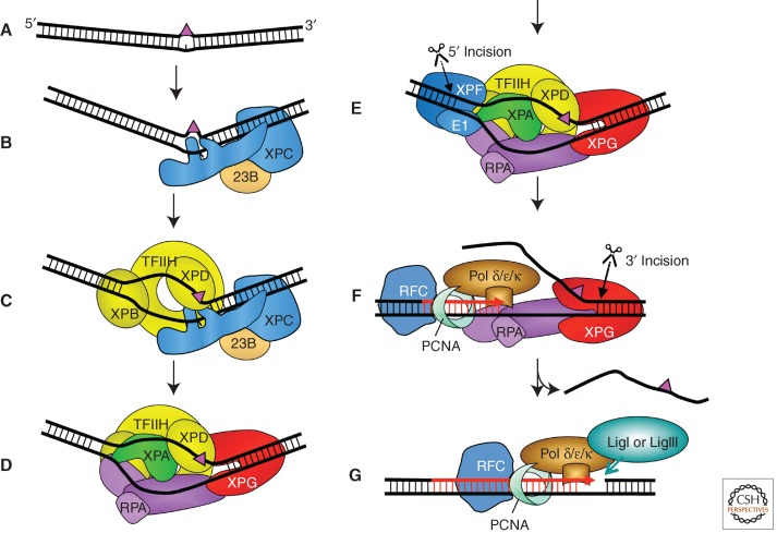
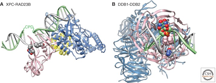
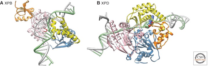
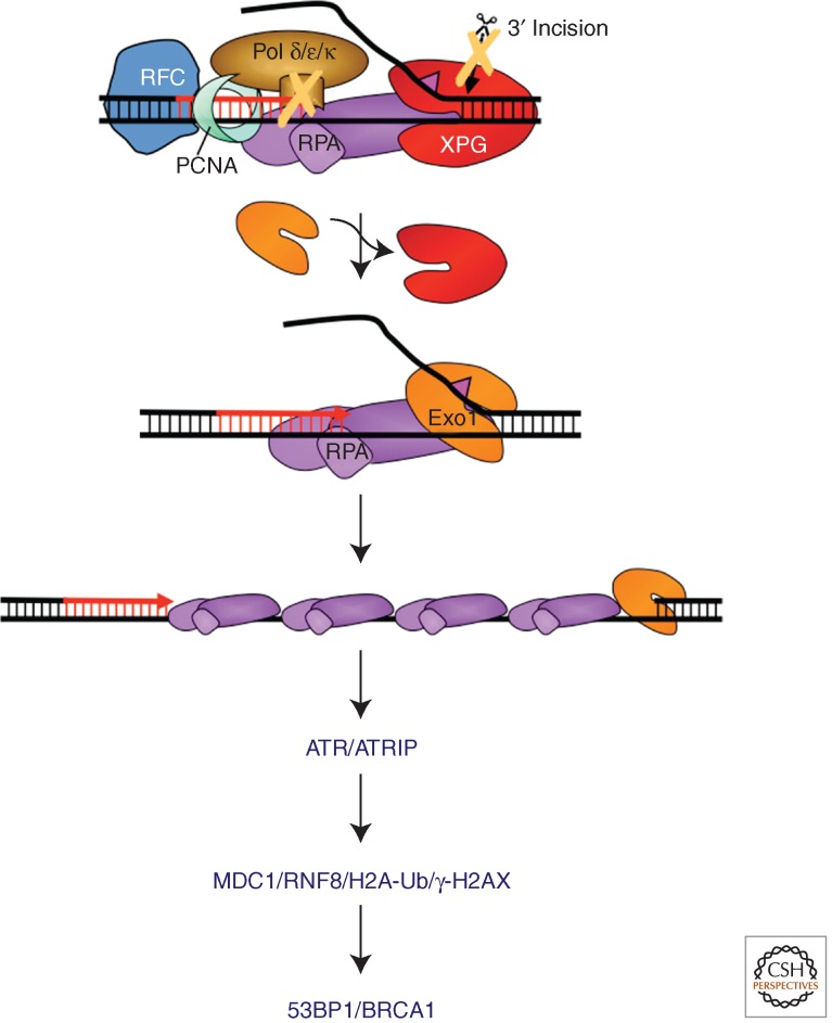
Nucleotide excision repair (NER) is the main pathway used by mammals to remove bulky DNA lesions such as those formed by UV light, environmental mutagens, and some cancer chemotherapeutic adducts from DNA. Deficiencies in NER are associated with the extremely skin cancer-prone inherited disorder xeroderma pigmentosum. Although the core NER reaction and the factors that execute it have been known for some years, recent studies have led to a much more detailed understanding of the NER mechanism, how NER operates in the context of chromatin, and how it is connected to other cellular processes such as DNA damage signaling and transcription. This review emphasizes biochemical, structural, cell biological, and genetic studies since 2005 that have shed light on many aspects of the NER pathway.


Figures

Figure 1.
Figure 1.
Model for the core NER reaction. (A) Bulky DNA lesions that destabilize duplex DNA are induced by a number of damaging agents. (B) In global genome NER, strongly distorting lesions are directly recognized by XPC-RAD23B, which binds the nondamaged strand opposite the lesion. (C) TFIIH interacts with XPC-RAD23B and pries the DNA open with its XPB subunit allowing XPD to track along DNA until stalls at the damage and verifies the chemical modification (bulkiness) of the lesion. (D) Stalling of XPD at the lesion allows for the formation of the preincision complex by recruitment of XPA, RPA, and XPG. The endonuclease XPG does not make an incision at this point. (E) Recruitment of ERCC1-XPF by interaction with XPA to the complex leads to incision 5′ to the lesion. (F) Initiation of repair synthesis by Pol δ and Pol κ or Pol ε and associated factors, followed by 3′ incision by XPG. (G) Completion of repair synthesis and sealing of the nick by DNA ligase IIIα/XRCC1 or DNA ligase I completes the process.
Figure 2.
Figure 2.
Structural basis for damage recognition in NER. (A) Structure of RAD4(XPC)-RAD23 bound to a CPD lesion. The TDG/BHD1 domains (blue) of RAD4/XPC bind to undamaged DNA, the BHD2/BHD3 domains (pink) encircle two nucleotides in the nondamaged strand of DNA. DNA is represented in gray with the two thymidine residues opposite the lesion in atom color. The position of the CPD lesion that is disordered in this structure is indicated. A fragment of RAD23 is shown in yellow. Note that the CPD lesion is present in a two base pair mismatch in this structure. The damaged DNA strand is shown in green, the undamaged strand in gray. The figure was made using the Chimera extensible molecular modeling system located at UCSF (www.cgl.ucsf.edu/chimera), using the structure PDB 2QSG (Min and Pavletich 2007). (B) Structure of DDB1-DDB2 bound to a CPD lesion. The UV-DDB complex, made up of DDB1 (light blue) and DDB2 (pink), binds DNA (gray) containing a CPD (atom color, green) through the DDB2 subunit. The DNA-binding site in DDB2 is located on one site of the β-propeller; DDB1 binds DDB2 on the opposite side. A wedge made up of residues F371, Q372, and H373 in DDB2 (shown in pink van der Waals representation) inserts into the DNA helix at the lesion site from the minor groove. The two modified nucleotides of the 6-4PP insert into a shallow binding pocket in DDB2. The damaged DNA strand is shown in green, the undamaged strand in gray. The figure was made using the Chimera extensible molecular modeling system located at UCSF (www.cgl.ucsf.edu/chimera), using the structure PDB A408 (Fischer et al. 2011).
Figure 3.
Figure 3.
Structural basis for helix opening and lesion verification in NER. (A) Model of an archaeal homolog of XPB bound to DNA. DNA-bound XPB undergoes a conformational change with respect to the apo protein, resulting in the insertion of the RED motif (red) into the duplex and binding of the DNA by the thumb domain (yellow). The two helicase domains are shown in pink (HD1) and blue (HD2), the damage-recognition domain (orange) is related to a similar domain found in MutS and aids initial DNA binding. (The figure was made using Chimera [www.cgl.ucsf.edu/chimera], based on data from Fan et al. 2006.) (B) Model of an archaeal XPD homolog tracking along DNA. The two helicase domains (HD1, blue; HD2, pink) bind the DNA at the ss/dsDNA (double-stranded DNA) junction, the FeS domain (orange, FeS cluster shown in yellow/red) and the arch domain encircle the ssDNA forming a tunnel through which the ssDNA translocate. Note that the tunnel is too narrow to permit bulky lesions to cross it, leading to stalling of the helicase and damage verification. Two native DNA bases in the tunnel are shown in green/atom color. (The figure was made using the Chimera extensible molecular modeling system located at UCSF [www.cgl.ucsf.edu/chimera], based on data from Fan et al. 2008.)
Figure 4.
Figure 4.
Model for NER-mediated UV-induced DNA-damage signaling. Damage signaling can be triggered during the repair synthesis step of NER if (1) the damage load is too high, (2) repair synthesis is inhibited, and (3) 3′ incision does not occur. In these situations, XPG is replaced with EXO1, which processed the NER intermediate into an ssDNA gap of up to kb in length. RPA covers the ssDNA and binds ATRIP/ATR activating the ATR kinase and triggering a signal cascade that involves MDC1, RNF8, 53BP1, and BRCA1, and leads to the establishment of chromatin marks, including ubiquitination of histones and phosphorylation of γ-H2AX. This process does not share many common features with DSB-induced signaling.

References

    1. Aboussekhra A, Biggerstaff M, Shivji MK, Vilpo JA, Moncollin V, Podust VN, Protic M, Hubscher U, Egly JM, Wood RD 1995. Mammalian DNA nucleotide excision repair reconstituted with purified protein components. Cell 80: 859–868 - PubMed
    1. Araujo SJ, Tirode F, Coin F, Pospiech H, Syvaoja JE, Stucki M, Hubscher U, Egly JM, Wood RD 2000. Nucleotide excision repair of DNA with recombinant human proteins: Definition of the minimal set of factors, active forms of TFIIH, and modulation by CAK. Genes Dev 14: 349–359 - PMC - PubMed
    1. Araujo SJ, Nigg EA, Wood RD 2001. Strong functional interactions of TFIIH with XPC and XPG in human DNA nucleotide excision repair, without a preassembled repairosome. Mol Cell Biol 21: 2281–2291 - PMC - PubMed
    1. Bergink S, Salomons FA, Hoogstraten D, Groothuis TA, de Waard H, Wu J, Yuan L, Citterio E, Houtsmuller AB, Neefjes J, et al. 2006. DNA damage triggers nucleotide excision repair-dependent monoubiquitylation of histone H2A. Genes Dev 20: 1343–1352 - PMC - PubMed
    1. Bergink S, Toussaint W, Luijsterburg MS, Dinant C, Alekseev S, Hoeijmakers JH, Dantuma NP, Houtsmuller AB, Vermeulen W 2012. Recognition of DNA damage by XPC coincides with disruption of the XPC-RAD23 complex. J Cell Biol 196: 681–688 - PMC - PubMed

Publication types

LinkOut - more resources