Review
. 2013 Oct 1;5(10):a012732.
doi: 10.1101/cshperspect.a012732.

Advances in understanding the complex mechanisms of DNA interstrand cross-link repair

Affiliations
Review

Advances in understanding the complex mechanisms of DNA interstrand cross-link repair

Cheryl Clauson et al. Cold Spring Harb Perspect Biol. .

Abstract

DNA interstrand cross-links (ICLs) are lesions caused by a variety of endogenous metabolites, environmental exposures, and cancer chemotherapeutic agents that have two reactive groups. The common feature of these diverse lesions is that two nucleotides on opposite strands are covalently joined. ICLs prevent the separation of two DNA strands and therefore essential cellular processes including DNA replication and transcription. ICLs are mainly detected in S phase when a replication fork stalls at an ICL. Damage signaling and repair of ICLs are promoted by the Fanconi anemia pathway and numerous posttranslational modifications of DNA repair and chromatin structural proteins. ICLs are also detected and repaired in nonreplicating cells, although the mechanism is less clear. A unique feature of ICL repair is that both strands of DNA must be incised to completely remove the lesion. This is accomplished in sequential steps to prevent creating multiple double-strand breaks. Unhooking of an ICL from one strand is followed by translesion synthesis to fill the gap and create an intact duplex DNA, harboring a remnant of the ICL. Removal of the lesion from the second strand is likely accomplished by nucleotide excision repair. Inadequate repair of ICLs is particularly detrimental to rapidly dividing cells, explaining the bone marrow failure characteristic of Fanconi anemia and why cross-linking agents are efficacious in cancer therapy. Herein, recent advances in our understanding of ICLs and the biological responses they trigger are discussed.


Figures
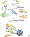
Figure 1.
Figure 1.
(A) Overview of the important steps in Fanconi anemia (FA) signaling pathway. Damage signaling begins with the recruitment of FANCM, FAAP24, and MHF to a stalled replication fork, binding to the unwound DNA. Remodeling of the fork by FANCM leads to recruitment of RPA, the ssDNA-binding protein. RPA localization to the DNA is required for ATR activation, which phosphorylates several targets, including the components of the MRN complex, FANCD2, and FANCI. The MRN complex associates with CtIP, which assists in DNA end resection during HR. The FA core complex assembles and includes FANCA, FANCB, FANCC, FANCE, FANCF, FANCG, FANCL, and FANCM, FAAP20, FAAP24, and MHF, using FANCM, FAAP24, and MHF to bind DNA. Assembly of the core complex stimulates FANCL to monoubiquitinate FANCD2 and FANCI. Structure-specific endonucleases: MUS81-EME1 and ERCC1-XPF/FANCQ are recruited to the damage site via interaction with FANCP/SLX4. The core complex and FANCD2-FANCI are recruited to the chromatin where they facilitate resolution of the repair intermediate by TLS polymerases (REV1- Pol ζ-Pol η) and the homologous recombination machinery, including FANCJ/BPIP1/BACH1, FANCD1/BRCA2, RAD51, FANCN/PALB2, FANCO/RAD51C, and BRCA1. (B) Details of the protein–protein interactions important for recruitment of FA proteins to sites of DNA damage. The core complex is recruited to chromatin via interaction of FAAP20 with RNF8 and the FANCC interaction with BRCA2/FANCD1. RNF8-UBC13 polyubiquitinates histone H2A, marking the site of ICL damage. FANCD2 and FANCI are recruited to chromatin via interaction between FANCD2 and FANCE of the core complex. FAAP20 also interacts with the TLS polymerase REV1. Dissolution of the complex is dependent on UAF1/USP1 deubiquitinating FANCD2 and FANCI. (C) Events that occur independently of the FA core complex. Recruitment of FANCM, MHF, and FAAP24 to stalled forks does not require the core complex to be present or monoubiquitination of FANCD2 or FANCI. In addition, RPA loading and ATR activation do not require the core complex members. Finally, homologous recombination, in particular the formation of 53BP1 and BRCA1 foci, FANCJ recruitment to the chromatin, DSB end resection by MRN-CtIP, and RAD51 loading on the resected end, do not require the core complex.
Figure 1.
Figure 1.
(A) Overview of the important steps in Fanconi anemia (FA) signaling pathway. Damage signaling begins with the recruitment of FANCM, FAAP24, and MHF to a stalled replication fork, binding to the unwound DNA. Remodeling of the fork by FANCM leads to recruitment of RPA, the ssDNA-binding protein. RPA localization to the DNA is required for ATR activation, which phosphorylates several targets, including the components of the MRN complex, FANCD2, and FANCI. The MRN complex associates with CtIP, which assists in DNA end resection during HR. The FA core complex assembles and includes FANCA, FANCB, FANCC, FANCE, FANCF, FANCG, FANCL, and FANCM, FAAP20, FAAP24, and MHF, using FANCM, FAAP24, and MHF to bind DNA. Assembly of the core complex stimulates FANCL to monoubiquitinate FANCD2 and FANCI. Structure-specific endonucleases: MUS81-EME1 and ERCC1-XPF/FANCQ are recruited to the damage site via interaction with FANCP/SLX4. The core complex and FANCD2-FANCI are recruited to the chromatin where they facilitate resolution of the repair intermediate by TLS polymerases (REV1- Pol ζ-Pol η) and the homologous recombination machinery, including FANCJ/BPIP1/BACH1, FANCD1/BRCA2, RAD51, FANCN/PALB2, FANCO/RAD51C, and BRCA1. (B) Details of the protein–protein interactions important for recruitment of FA proteins to sites of DNA damage. The core complex is recruited to chromatin via interaction of FAAP20 with RNF8 and the FANCC interaction with BRCA2/FANCD1. RNF8-UBC13 polyubiquitinates histone H2A, marking the site of ICL damage. FANCD2 and FANCI are recruited to chromatin via interaction between FANCD2 and FANCE of the core complex. FAAP20 also interacts with the TLS polymerase REV1. Dissolution of the complex is dependent on UAF1/USP1 deubiquitinating FANCD2 and FANCI. (C) Events that occur independently of the FA core complex. Recruitment of FANCM, MHF, and FAAP24 to stalled forks does not require the core complex to be present or monoubiquitination of FANCD2 or FANCI. In addition, RPA loading and ATR activation do not require the core complex members. Finally, homologous recombination, in particular the formation of 53BP1 and BRCA1 foci, FANCJ recruitment to the chromatin, DSB end resection by MRN-CtIP, and RAD51 loading on the resected end, do not require the core complex.
Figure 2.
Figure 2.
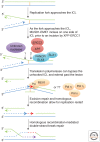
Current model for replication-dependent ICL repair. As a replication fork approaches an ICL, the fork stalls 20–40 nucleotides from the lesion. Two incisions are made on the lagging strand template. The first incision creates a single-ended double-strand break. The identity of the nuclease making this incision is currently not known. Then FANCQ/XPF-ERCC1 completely unhooks the ICL from the lagging strand template with a second nick. Both endonucleases are recruited to the damage site by FANCP/SLX4. Now translesion polymerases are able to bypass the unhooked ICL using the leading strand as a primer. Different TLS polymerases may be required to bypass various unhooked ICLs, whereas Pol ζ-REV1 is adept at extending mismatches created by bypass insertion. This DNA synthesis is required to enable homologous recombination-mediated repair of the broken end.
Figure 3.
Figure 3.
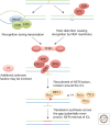
Current model for replication-independent ICL repair. If an ICL occurs in nonreplicating cells, it may be recognized by interfering with transcription or because it induces helix distortion, which is recognized by the nucleotide excision repair pathway. This could trigger recruitment of downstream NER factors including XPF-ERCC1. Incisions around the lesion on one strand of DNA unhook the lesion from that strand. Translesion polymerases can bypass the ICL and fill the gap with new DNA synthesis. This is sufficient to restore double-strand DNA that is free of interstrand cross-links.

References

    1. Abraham J, Lemmers B, Hande MP, Moynahan ME, Chahwan C, Ciccia A, Essers J, Hanada K, Chahwan R, Khaw AK, et al. 2003. Eme1 is involved in DNA damage processing and maintenance of genomic stability in mammalian cells. EMBO J 22: 6137–6147. - PMC - PubMed
    1. Albertella MR, Green CM, Lehmann AR, O’Connor MJ. 2005. A role for polymerase η in the cellular tolerance to cisplatin-induced damage. Cancer Res 65: 9799–9806. - PubMed
    1. Al-Minawi AZ, Lee Y-F, Håkansson D, Johansson F, Lundin C, Saleh-Gohari N, Schultz N, Jenssen D, Bryant HE, Meuth M, et al. 2009. The ERCC1/XPF endonuclease is required for completion of homologous recombination at DNA replication forks stalled by inter-strand cross-links. Nucleic Acids Res 37: 6400–6413. - PMC - PubMed
    1. Andersen SL, Bergstralh DT, Kohl KP, LaRocque JR, Moore CB, Sekelsky J. 2009. Drosophila MUS312 and the vertebrate ortholog BTBD12 interact with DNA structure-specific endonucleases in DNA repair and recombination. Mol Cell 35: 128–135. - PMC - PubMed
    1. Andreassen PR, D’Andrea AD, Taniguchi T. 2004. ATR couples FANCD2 monoubiquitination to the DNA-damage response. Genes Dev 18: 1958–1963. - PMC - PubMed

MeSH terms

LinkOut - more resources