Review
. 2013 Oct 1;3(10):a013847.
doi: 10.1101/cshperspect.a013847.

Embryonic heart progenitors and cardiogenesis

Affiliations
Review

Embryonic heart progenitors and cardiogenesis

Thomas Brade et al. Cold Spring Harb Perspect Med. .

Abstract

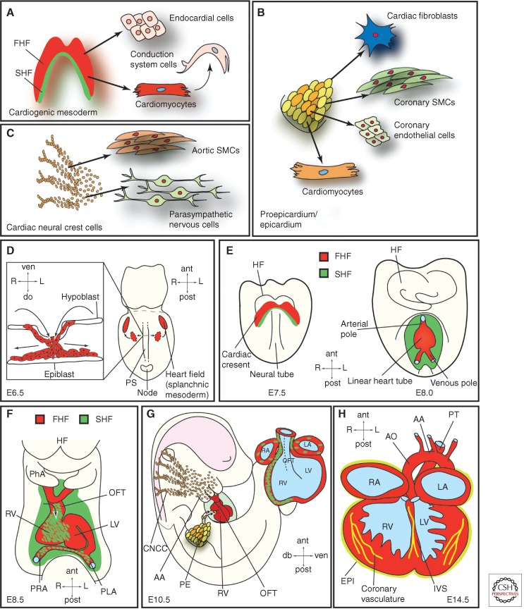
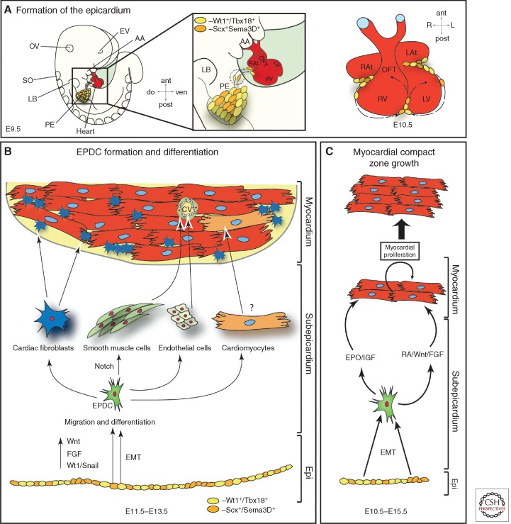
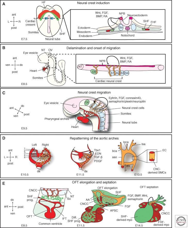
The mammalian heart is a highly specialized organ, comprised of many different cell types arising from distinct embryonic progenitor populations during cardiogenesis. Three precursor populations have been identified to contribute to different myocytic and nonmyocytic cell lineages of the heart: cardiogenic mesoderm cells (CMC), the proepicardium (PE), and cardiac neural crest cells (CNCCs). This review will focus on molecular cues necessary for proper induction, expansion, and lineage-specific differentiation of these progenitor populations during cardiac development in vivo. Moreover, we will briefly discuss how the knowledge gained on embryonic heart progenitor biology can be used to develop novel therapeutic strategies for the management of congenital heart disease as well as for improvement of cardiac function in ischemic heart disease.


Figures

Figure 1.
Figure 1.
Embryonic heart progenitor contributions to different cardiac compartments and cell types during heart morphogenesis in mouse development. (A) Cardiogenic mesoderm, (B) proepicardium/epicardium, and (C) cardiac neural crest cell lineage diversification. (D) At E6.5, mesodermal progenitors ingress through the primitive streak (PS) and migrate away from the PS (illustrated in the box showing a transverse section) to form the heart fields located in the splanchnic mesoderm. (E) At E7.5, the first and second heart fields (FHF and SHF) are discernible. The first heart fields fuse at the midline thereby forming the cardiac crescent caudal to the headfolds. FHF progenitor cells start to differentiate. At E8.0, the cardiac crescent forms the beating, linear heart tube. SHF progenitors will gradually migrate into the linear heart tube and differentiate then. (F) At E8.5, the linear heart tube undergoes rightward looping. (G) At E10.5, cardiac neural crest and proepicardial cells contribute to the heart, which already shows a defined four-chamber morphology. (H) At E14.5, the heart shows four fully septated chambers and a septated outflow tract connected to the pulmonary trunk and the dorsal aorta. AA, Aortic arch; ant, anterior; AO, dorsal aorta; CNCC, cardiac neural crest cells; do, dorsal; EPI, epicardium; FHF, first heart field; HF, headfolds; IVS, interventricular septum; L, left; LA, left atrium; LV, left ventricle; OFT, outflow tract; PE, proepicardium; PhA, pharyngeal arch; PLA, primitive left atrium; post, posterior; PRA, primitive right atrium; PS, primitive streak; PT, pulmonary trunk; R, right; RA, right atrium; RV, right ventricle; SHF, second heart field; SMCs, smooth muscle cells; ven, ventral.
Figure 2.
Figure 2.
Cellular hierarchy of cardiac progenitor cells and their lineage specification. Several signaling pathways (BMP, Nodal, Wnt/β-catenin, FGF) interact to induce mesoderm; Brachyury (Bry) positive mesodermal precursors first differentiate through Bry+/Flk-1+ hemangioblasts toward endothelial and blood-cell lineages (around E5.5 during mouse development). Slightly later during development after down-regulation of Wnt/β-catenin signaling and induction of noncannonical Wnt signals a second wave of Bry+/Flk-1+ mesodermal progenitors appears. Eomesodermin signaling drives cardiogenic mesoderm specification from these primitive mesodermal precursors. Cardiogenic mesoderm is marked by the expression of mesoderm posterior 1 (Mesp1) (around E6.5 in mouse embryogenesis). Early mesoderm-derived cardiac precursors undergo further lineage restriction and differentiate into progenitor pools that populate the FHF and SHF, respectively. At this stage (E7.5 mouse development) FHF progenitors start to differentiate upon BMP and FGF action toward cardiomyocytes and smooth muscle cells, whereas Wnt/β-catenin, FGF, and endodermal Shh signaling keeps SHF progenitors in a proliferative state. These SHF progenitors are defined by the molecular signature Isl-1+/Nkx2.5+/Flk-1+. SHF progenitors are now gradually added to the looping heart tube and get further restricted in their differentiation potential (E8.5). Two subpopulations of SHF progenitors can be distinguished. One population marked by the expression of Isl-1 and Flk-1 differentiates into endothelial cells and smooth muscle cells, whereas a second pool of Isl-1+/Nkx2.5+ SHF precursors provides smooth muscle cells and cardiomyocytes as well as contributing to the proepicardial lineages (Wt1+/Tbx18+ and Scx+/Sema3D+ populations), which later form cardiac fibroblasts (CF), smooth muscle cells (SMCs), endothelial cells (EC), and cardiomyocytes (CM), with the latter contribution being still unclear. These distinct SHF progenitor populations differentiate upon BMP signals from the lateral plate mesoderm as well as Notch and noncanonical Wnt signals. SHF patterning is governed by RA and TGF-β signals.
Figure 3.
Figure 3.
Origin and fates of the proepicardium. (A) Formation of the epicardium. Starting around E9.5 of mouse development, freely floating proepicardial cell vesicles attach to the naked myocardium, starting in the atrioventricular canal region. The whole heart will be covered by an epicardial epithelial sheet by E11.5. (B) Formation of epicardium-derived cells (EPDCs). At E11.5–13.5, epicardial cells undergo an epithelial-mesenchymal transition (EMT) (mediated by Wnt, FGF, and Wt1/Snail signals) and EPDCs emerge in the subepicardial space. EPDCs then differentiate toward various cell types, as indicated, and contribute to the coronary vasculature (cv) and cardiac fibroblast. EPDC differentiation toward the myocytic lineage is still under thorough investigation. (C) Epicardium/EPDCs are secreting important factors that induce myocardial proliferation and thereby myocardial compact zone growth. AA, Aortic arches; ant, anterior; cv, coronary vessels; do, dorsal; EMT, epithelial to mesenchymal transition; EPDC, epicardial-derived cells; Epi, epicardium; EPO, erythropoietin; EV, eye vesicle; IGF, insulin growth factor; L, left; LAt, left atrium; LB, limb bud; LV, left ventricle; OFT, outflow tract; OV, otic vesicle; PE, proepicardium; post, posterior; R, right; RA, retinoic acid; RAt, right atrium; RV, right ventricle; SO, somites; ven. ventral.
Figure 4.
Figure 4.
Cardiac neural crest. (A) Neural crest induction. Illustrated is a ventral view of an E7.5 embryo and a transverse section through the first somite pair. The section detail shows the different germ layers and factors originating in the ectoderm and mesoderm that are required for neural crest cell induction in the neural plate border (NPB). Ant, anterior; post, posterior; R, right; L, left; HF, headfolds; FHF, first heart field; SHF, second heart field; So, somite. (B) Delamination and onset of migration of cardiac neural crest cells. At E8.5, cardiac neural crest cells delaminate from the NPB between the mid-otic vesicle (ov) and the posterior border of somite 3 (S3) and start on their migratory path toward the heart. These processes are mediated by Wnt, FGF, BMP, and RA signals. Please see text for more details. ant, anterior; post, posterior; ven, ventral; do, dorsal; S1,2,3,4, somite 1,2,3,4. (C) Neural crest migration. By E9.5, the CNCCs reach the aortic arches and are proceeding toward the OFT. Signals targeting the CNCCs to the aortic arches are indicated. ov, otic vesicle. (D) The CNCCs are vital for the repatterning of the aortic arches. There are initially six symmetrical arteries attached to the parallel dorsal aorta (da). Sequentially these arteries are remodeled and give rise to a separate ascending aorta (aao) and pulmonary trunk (P) with two pulmonary arteries attached. Additionally, the cardiac neural crest cells are differentiating into aortic smooth muscle cells and envelope the nascent endothelial cells forming the aortic arches. Required factors for both processes are indicated (END, endothelin); see text for details. bca, Brachiochepalic artery; lcca, left common carotid artery; lsa, left subclavian artery. (E) Interaction of CNCCs and SHF progenitors leads to outflow tract (OFT) elongation and septation. At E11.5, cardiac neural crest cells condense and form the U-shaped aorticopulmonary septation complex (APSC); see text for details. CNCC, Cardiac neural crest cells; SHF prog., second heart field progenitors; NT, neural tube; So, somites; IFT, inflow tract; diff. SHF prog., differentiated second heart field progenitors; AA, aortic arches; FHF derived myo., first heart field-derived myocardium; SHF derived myo, second heart field-derived myocardium.

References

    1. Abu-Issa R, Smyth G, Smoak I, Yamamura K, Meyers EN 2002. Fgf8 is required for pharyngeal arch and cardiovascular development in the mouse. Development 129: 4613–4625 - PubMed
    1. Abu-Issa R, Waldo K, Kirby ML 2004. Heart fields: One, two or more? Dev Biol 272: 281–285 - PubMed
    1. Aguirre A, Sancho-Martinez I, Izpisua Belmonte JC 2013. Reprogramming toward heart regeneration: Stem cells and beyond. Cell Stem Cell 12: 275–284 - PubMed
    1. Arceci RJ, King AA, Simon MC, Orkin SH, Wilson DB 1993. Mouse GATA-4: A retinoic acid-inducible GATA-binding transcription factor expressed in endodermally derived tissues and heart. Mol Cell Biol 13: 2235–2246 - PMC - PubMed
    1. Bergmann O, Bhardwaj RD, Bernard S, Zdunek S, Barnabe-Heider F, Walsh S, Zupicich J, Alkass K, Buchholz BA, Druid H, et al. 2009. Evidence for cardiomyocyte renewal in humans. Science 324: 98–102 - PMC - PubMed

Publication types