Review
. 2013 Oct 1;3(10):a016717.
doi: 10.1101/cshperspect.a016717.

The inside story of Shigella invasion of intestinal epithelial cells

Affiliations
Review

The inside story of Shigella invasion of intestinal epithelial cells

Nathalie Carayol et al. Cold Spring Harb Perspect Med. .

Abstract

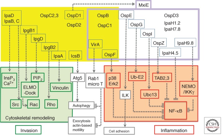
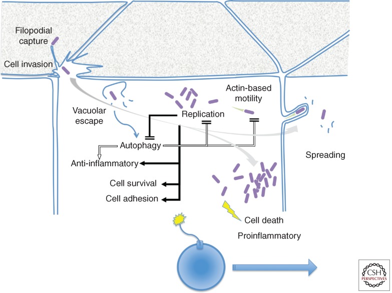
As opposed to other invasive pathogens that reside into host cells in a parasitic mode, Shigella, the causative agent of bacillary dysentery, invades the colonic mucosa but does not penetrate further to survive into deeper tissues. Instead, Shigella invades, replicates, and disseminates within the colonic mucosa. Bacterial invasion and spreading in intestinal epithelium lead to the elicitation of inflammatory responses responsible for the tissue destruction and shedding in the environment for further infection of other hosts. In this article, we highlight specific features of the Shigella arsenal of virulence determinants injected by a type III secretion apparatus (T3SA) that point to the targeting of intestinal epithelial cells as a discrete route of invasion during the initial event of the infectious process.


Figures

Figure 1.
Figure 1.
The control of cell responses by Shigella T3S effectors. Shigella T3S effectors are shown in yellow and purple boxes. The T3S effectors boxed in blue represent those for which a function has been assigned. T3S effectors that are constitutively expressed (yellow box) or up-regulated following cell-contact induction of secretion (boxed in purple) and activation by MxiE. On cell contact, IpaB and IpaC insert into the host-cell membrane to form the “translocon,” required for the induction of InsP3-dependent Ca2+ responses. The targets of T3S effectors involved in Shigella invasion are boxed in green. IpaC, through its carboxy-terminal domain induces the recruitment and activation of the Src kinase. IpgB1 and IpgD, together with IpaC, participate in actin polymerization and membrane ruffle formation by targeting ELMO/Dock and hydrolyzing PIP2, respectively. IpgB2 that activates Rho, and IpaA that binds to vinculin, further reorganize the actin cytoskeleton to promote invasion. IcsB prevents autophagic responses by inhibiting binding of Atg5 to IcsA. The targets of T3S effectors that down-regulate inflammation are boxed in red. VirA prevents autophagy and IL-8 secretion through its GAP activity toward Rab1. VirA also inhibits microtubule polymerization to favor actin-based motility. Among the T3S effectors up-regulated by MxiE, OspF prevents the activation of the p38 and Erk2 MAP kinases via its phospho-threonine lyase activity. OspE reinforces cell adhesion by targeting ILK. The targets of OspG, OspI, OspZ, IpaH4.5, and IpaH9.8 that down-regulate NF-κB activation are indicated.
Figure 2.
Figure 2.
Filopodial capture and Shigella invasion at the apical side of polarized Caco-2 cells. Shown is a Z projection of confocal fluorescence images of Shigella-induced actin foci at the apical side of polarized Caco-2 cells. Red, anti-LPS immunostaining; green, Alexa488-phalloidin staining. Z sections were acquired via spinning-disk confocal microscopy. Scale bar, 5 mm.
Figure 3.
Figure 3.
Shigella invasion, intracellular replication, and spreading in epithelial cells. Filopodial capture initiates the invasion process, accompanied by localized membrane ruffles at the epithelial cell apical surface. Following invasion, bacterial escape from the vacuole triggers autophagic responses that dampen inflammatory signals and limit bacterial replication and actin-based motility. During the early stages of bacterial intracellular replication, T3S effectors counter autophagy, down-regulate inflammatory responses, promote cell survival, and reinforce cell adhesion (solid arrows) to preserve the epithelial cell integrity (grey arrow), to allow bacterial spreading. At late stages of intracellular replication, the increase in cytosolic Ca2+ concentration and mitochondrial damage lead to necrotic cell death and the emission of proinflammatory signals (yellow). Shigella invasion of epithelial cells can be seen as the ignition of a time bomb, in which bacterial spreading must occur before necrotic death of the primary infected cell.

References

    1. Alto NM, Shao F, Lazar CS, Brost RL, Chua G, Mattoo S, McMahon SA, Ghosh P, Hughes TR, Boone C, et al. 2006. Identification of a bacterial type III effector family with G protein mimicry functions. Cell 124: 133–145 - PubMed
    1. Arbibe L, Kim DW, Batsche E, Pedron T, Mateescu B, Muchardt C, Parsot C, Sansonetti PJ 2007. An injected bacterial effector targets chromatin access for transcription factor NF-κB to alter transcription of host genes involved in immune responses. Nat Immunol 8: 47–56 - PubMed
    1. Ashida H, Kim M, Schmidt-Supprian M, Ma A, Ogawa M, Sasakawa C 2010. A bacterial E3 ubiquitin ligase IpaH9.8 targets NEMO/IKKγ to dampen the host NF-κB-mediated inflammatory response. Nat Cell Biol 12: 66–73 - PMC - PubMed
    1. Ashida H, Ogawa M, Kim M, Suzuki S, Sanada T, Punginelli C, Mimuro H, Sasakawa C 2011. Shigella deploys multiple countermeasures against host innate immune responses. Curr Opin Microbiol 14: 16–23 - PubMed
    1. Bergounioux J, Elisee R, Prunier AL, Donnadieu F, Sperandio B, Sansonetti P, Arbibe L 2012. Calpain activation by the Shigella flexneri effector VirA regulates key steps in the formation and life of the bacterium’s epithelial niche. Cell Host Microbe 11: 240–252 - PubMed

Publication types

MeSH terms

Substances

LinkOut - more resources