. 2013 Sep;11(9):e1001666.
doi: 10.1371/journal.pbio.1001666. Epub 2013 Sep 24.

Hypoxic regulation of hand1 controls the fetal-neonatal switch in cardiac metabolism

Affiliations

Hypoxic regulation of hand1 controls the fetal-neonatal switch in cardiac metabolism

Ross A Breckenridge et al. PLoS Biol. 2013 Sep.

Erratum in

  • PLoS Biol. 2013 Dec;11(12). doi:10.1371/annotation/a9a7f37a-3fa7-4f7f-8310-1339bf5a666e

Abstract

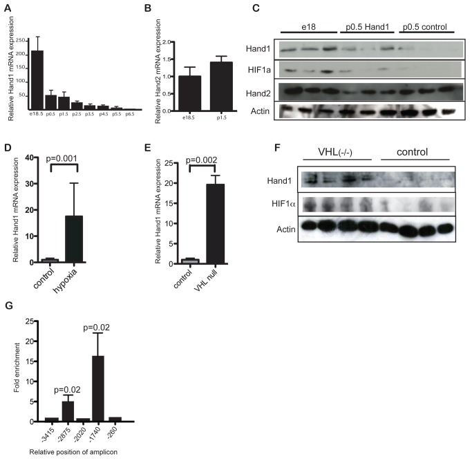
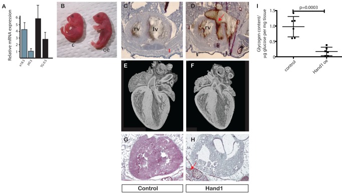
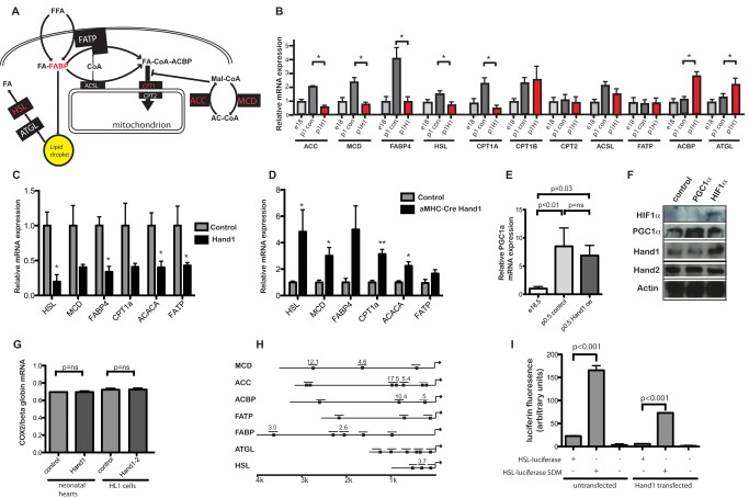
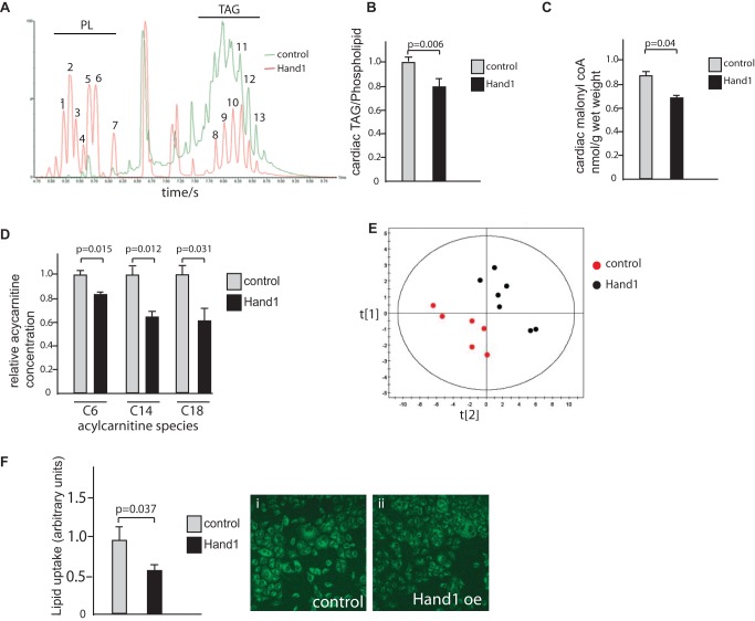
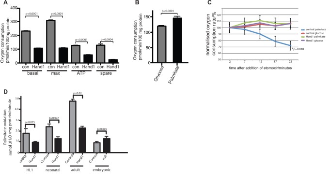
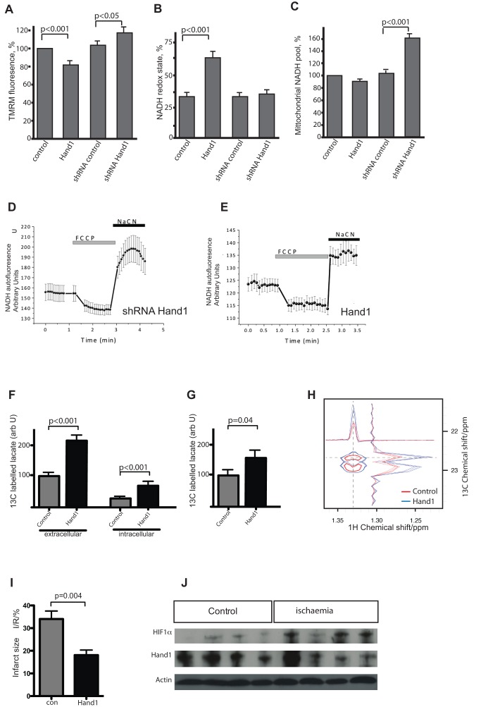
Cardiomyocytes are vulnerable to hypoxia in the adult, but adapted to hypoxia in utero. Current understanding of endogenous cardiac oxygen sensing pathways is limited. Myocardial oxygen consumption is determined by regulation of energy metabolism, which shifts from glycolysis to lipid oxidation soon after birth, and is reversed in failing adult hearts, accompanying re-expression of several "fetal" genes whose role in disease phenotypes remains unknown. Here we show that hypoxia-controlled expression of the transcription factor Hand1 determines oxygen consumption by inhibition of lipid metabolism in the fetal and adult cardiomyocyte, leading to downregulation of mitochondrial energy generation. Hand1 is under direct transcriptional control by HIF1α. Transgenic mice prolonging cardiac Hand1 expression die immediately following birth, failing to activate the neonatal lipid metabolising gene expression programme. Deletion of Hand1 in embryonic cardiomyocytes results in premature expression of these genes. Using metabolic flux analysis, we show that Hand1 expression controls cardiomyocyte oxygen consumption by direct transcriptional repression of lipid metabolising genes. This leads, in turn, to increased production of lactate from glucose, decreased lipid oxidation, reduced inner mitochondrial membrane potential, and mitochondrial ATP generation. We found that this pathway is active in adult cardiomyocytes. Up-regulation of Hand1 is protective in a mouse model of myocardial ischaemia. We propose that Hand1 is part of a novel regulatory pathway linking cardiac oxygen levels with oxygen consumption. Understanding hypoxia adaptation in the fetal heart may allow development of strategies to protect cardiomyocytes vulnerable to ischaemia, for example during cardiac ischaemia or surgery.


Conflict of interest statement

The authors have declared that no competing interests exist.

Figures

Figure 1
Figure 1. Hand1 levels fall in the heart immediately following birth, under control of hypoxia signaling.
(A) RTPCR for Hand1 RNA from whole hearts of perinatal mice at a range of stages around birth, showing a steep decline in expression from birth. Levels expressed as a multiple of average 6-wk-old adult levels (n = 4 each group). (B) Levels of cardiac Hand2 RNA do not fall at birth. Levels of Hand2 at p1.5 normalised to e18.5 levels (n = 6 each group). (C) Western blot of protein extract from e18 (prenatal) control, p0.5 XMLC-Hand1, and p0.5 control hearts showing reduction in Hand1 but not Hand2 protein levels after birth, and persistence of Hand1 expression in XMLC2-Hand1 hearts. (D) RTPCR showing increased Hand1 RNA levels in hearts of adult wild-type mice incubated at 12% oxygen for 2 wk (“hypoxia”) over controls at normoxia (20% O2) (n = 4 each group) (p = 0.001, two-tailed t test). (E) RTPCR showing significantly increased Hand1 RNA levels in the hearts of p0.5 neonatal α-MHC-Cre::VHL(fl/fl ) mice compared with wild-type controls, p = 0.0002 two-tailed t test, n = 6 each group. (F) Western blot of protein extract from VHL(fl/fl) and control hearts at p0.5, showing elevation of Hand1 and HIF1α in VHL(fl/fl) hearts. (G) RTPCR of chromatin immunoprecipitation assay using anti-HIF1α antiserum and primers to the HIF motif-containing sequences in the Hand1 promoter from e18 hearts, showing binding of HIF1α to two sites. Bars represent summation of three experiments, and results expressed as multiples of signal for nonamplified sequence. The p values are two-tailed t tests relative to nonamplified γ-crystallin primers.
Figure 2
Figure 2. Prevention of neonatal Hand1 down-regulation in transgenic mouse hearts leads to cardiomyopathy and death.
(A) Cardiac RNA levels of Hand1 in e18.5 and p0.5 wild-type, and e18.5 and p0.5 transgenic (TG 18.5 and TG0.5, respectively) showing RNA levels in the transgenic heart around 2.5 times that of wild-type p0.5 heart. (B) Cardiac Hand1 elevating pups appear grossly normal, but are cyanosed (c, control; oe, Hand1 overexpressing). (C, D) H and E stain of cryostat section through thorax of control and Hand1 overexpressing hearts, showing thin ventricular wall of the Hand1 overexpressing heart, and ventricular rupture (arrowed) with blood in the pericardial space (rv, right ventricle; lv, left ventricle). (E, F) EFIC sectioning and reconstruction of control and Hand1 overexpressing hearts from 4-h-old fostered pups, showing small size but no gross structural defect. (G, H) Periodic acid-Schiff stain of control and Hand1 overexpressing heart, showing decreased glycogen levels in Hand1 overexpressing heart (purple). Glycogen stain in intercostal muscle of transgenic pup arrowed in (H). (I) Quantification of glucose enzymatically released from glycogen in hearts of neonates 2 h after caesarian section. Levels of glycogen in XMLC-Hand1 hearts are 17.5% of XMLC controls (p value, two-tailed t test, n = 6 hearts each group).
Figure 3
Figure 3. Prolongation of neonatal cardiac Hand1 expression prevents transcriptional up-regulation of lipid metabolizing genes.
(A) Schematic showing myocardial lipid metabolism (adapted from Kodde et al. [55]). (B) RTPCR showing RNA expression in e16 control, p0.5 control, and p0.5 hand1 up-regulating hearts. Levels of ACC, MCD, FABP4, ACSL, CPT1A CPT1B, and HSL are significantly up-regulated around birth (p<0.05 two-tailed t test, n = 4 each group). No postnatal rise in ACC, MCD, FABP4, CPT1A, and HSL is seen in Hand1 up-regulating hearts. Significantly increased RNA expression of ACBP and ATGL is seen in Hand1 up-regulating hearts. Genes whose expression is reduced in Hand1 overexpressing hearts are in red in (A). ACC, acetyl coA carboxylase; MCD, malonyl coA decarboxylase; FABP, fatty acid binding protein; FATP, fatty acid transport protein; ACSL, acyl coA synthase long chain 1; HSL, hormone sensitive lipase; ATGL, adipose triglyceride lipase; ACBP, acylcoA binding protein; CPT, Carnitine Palmitoyl Transferase. (C) RTPCR of mRNA from 2-mo-old adult XMLC2-Hand1 mice following doxycycline induction for 2 wk, showing changes in expression of RNA encoding fatty acid metabolising proteins relative to control non-up-regulating mice (*p<0.05, **p<0.005, two-tailed t test, n = 4 each group). (D) RTPCR of mRNA from e14.5 embryo hearts from αMHC-Cre::Hand1(fl/fl) and control pups, showing up-regulation of genes encoding fatty acid metabolising enzymes(*p<0.05, two-tailed t test, n = 4 each group). (E) RTPCR showing significant PGC1-α elevation in the heart around birth (n = 4 each group, p = 0.008, two-tailed t test), with no significant drop in Hand1 up-regulating hearts (p = 0.26, n = 6 each group). (F) Western blot of protein extract from cultured HL1 cardiomyocytes nontransfected (“control”) and transfected with PGC1-α and HIF1, showing no elevation of Hand1 in PGC1-α elevated PGC1α and Hand1 but not Hand2 protein expression in HIF1 expressing cells. (G) PCR of nuclear genomic (globin) and mitochondrial (COX2) DNA showing unchanged ratio in Hand1 elevating neonatal hearts and Hand1-transfected HL1 cells compared with controls, implying no change in mitochondrial number. Control HL1 cells are transfected with an empty vector. (H) Map of 5′ promoters of several putative Hand1 transcriptional targets in the e18.5 heart. Numbers refer to fold enrichment over γ crystallin in chromatin immunoprecipitation assay using anti-Hand1 serum. For more detailed chromatin immunoprecipitation data, please see Text S1 and Figure S2. (I) Site-directed mutagenesis of the Hand1-binding canonical CANNTG e-box in the 5′ HSL luciferase promoter de-represses expression of luciferase in HL1 cells, both in untransfected cells and cells stably expressing Hand1 (transfections in triplicate, measurement in quadruplicate, p values, two-tailed t test).
Figure 4
Figure 4. Lipid metabolism is inhibited in neonatal Hand1 overexpressing hearts.
(A) LC-MS trace showing typical output for intact lipid extracted from control hearts (green trace) and Hand1 up-regulating hearts (red), showing significantly lower levels of triacylglycerides (TAG) compared to phospholipid (PL) in Hand1 up-regulating hearts. (B) Quantitative analysis of cardiac triacylglyceride levels showing significant reduction in Hand1 up-regulating hearts, expressed as the ratio of TAG to phospholipid (n = 6 each group, p = 0.006, two-tailed t test). (C) Quantitative analysis of cardiac malonyl coA levels showing significant reduction in Hand1 up-regulating hearts (n = 6 each group, p = 0.04, two-tailed t test). (D) Reduced levels of C6, C14, and C18 containing acylcarnitine species in Hand1 prolonging neonatal hearts compared with controls. For full dataset, please refer to Table S3. (E) Multivariate partial least squares discriminant analysis (PLS-DA) scores of acylcarnitine profiles showing a significant decrease in global levels of acylcarnitines in Hand1 up-regulated hearts relative to controls (R2X = 33%, R2Y = 62%, and Q2 = 48%). (F) BODIPY-500/510C1, C12 uptake is significantly reduced in HL1 cells by transfection with Hand1. Graph shows quantification of fluorescence in Hand1 transfected and nontransfected controls, 10 high power fields each. (i) and (ii) show representative fluorescence micrographs of control and Hand1 transfected cells following labeled lipid incubation (p = 0.037, two-tailed t test).
Figure 5
Figure 5. Hand1 reduces oxygen consumption and lipid oxidation in stably transfected HL1 cells and primary cardiomyocytes.
(A) In HL1 cells stably transfected with Hand1, basal respiration (basal) maximal respiratory capacity (max), ATP production (ATP), and spare respiratory capacity (spare) are all significantly reduced compared to empty-vector transfected controls, using medium that contains 5.5 mmol glucose and 2 mmol pyruvate (five duplicate wells for each measurement, experiments repeated three times, two-tailed t test). (B) Oxygen consumption is increased in HL1 cells following incubation with 20 mmol palmitate for 20 min compared with cells incubated in 12 mmol glucose (15 replicate wells, two-tailed t test). Substrate was added to basal incubation medium that contains 5.5 mmol glucose and 2 mmol pyruvate. (C) The CPT1 inhibitor etomoxir (1 µM) reduces oxygen consumption in nontransfected but not Hand1 expressing HL1 cells when incubated with 20 mM palmitate (p = 0.018 AUC ANOVA, 15 wells per sample). No reduction in oxygen consumption is seen in either cell type when cells incubated with 12 mM glucose are treated with etomoxir (i.e., undergoing glycolyic respiration). Measurements are carried out after treatment with oligomycin and FCCP. (D) Hand1 reduces palmitate oxidation in cardiomyocytes. HL1 cells stably expressing Hand1 generate significantly less 3H20 from 3H labeled palmitate compared with a line stably expressing an shRNA construct directed against Hand1, showing lower levels of lipid oxidation. Bars represent sum of three experiments (p = 0.015, two-tailed t test). Primary cultures of Hand1 up-regulating neonatal cardiomyocytes (four hearts each group) and adult hearts (two hearts each group) generate significantly less 3H20 than controls (two-tailed t test). Primary cultured cardiomyocytes from e15 Hand1null αMHC-Cre::Hand1(fl/fl) (three hearts each group) exhibit significantly increased levels of lipid uptake (two-tailed t test).
Figure 6
Figure 6. Hand1 levels control mitochondrial function, metabolic flux, and ischaemia susceptibility in cardiomyocytes.
(A) TMRM fluorescence measured in stably transfected HL1 cell lines, showing significantly reduced mitochondrial inner membrane potential in Hand1 up-regulation, and significantly increased potential in shRNA-expressing lines knocking down Hand1 expression (number of cells analysed = 26, 34, 23, and 23, respectively) (p = two-tailed t test). (B) Significantly increased NADH redox state in Hand1 expressing stable HL1 lines, measured by NADH autofluoresence. (C) Hand1 up-regulating stably transfected HL1 cells display a significantly reduced mitochondrial NADH pool compared with controls, whereas knockdown of Hand1 results in an increase in the NADH pool, estimated as a difference in fluorescence (arbitrary U) between responses to FCCP and NaCN. (D) Example of an NADH autofluoresence trace of a stably transfected Hand1 shRNA HL1 cell, showing maximally oxidized state in response to the uncoupler FCCP, and minimally oxidized state in response to sodium cyanide, in comparison to the trace for Hand1 transfected cell. (E) A typical NADH autofluoresence trace of a stably transfected Hand1 line, showing much lower NADH levels, as defined by the ratio of maximally and minimally oxidized states. (F) 13C labeling of lactate is increased in supernatant (extracellular) and cell extract (intracellular) of stably transfected Hand1 up-regulating HL1 cells after labeling of cells with uniformly labeled 13C glucose (n = 4 for each measurement), measured by 1H. (G) Increased detection of 13C labeling of lactate in adult XMLC-Hand1 mice 1 h after administration of uniformly labeled 13C glucose (n = 6 each group, p = 0.04, two-tailed t test). (H) 1H NMR showing increased 13C incorporation into lactate from [U 13C6]-glucose in Hand1 up-regulating HL1 cells (blue trace) compared to controls (red trace). (I) Following Langendorff perfusion and 35 min of global ischaemia followed by 30 min of reperfusion, hearts from 2-mo-old Hand1 up-regulating XMLCrTTA::tetHand1 adult mice following 1 mo of doxycycline induction exhibit a 47% reduction in infarct size, infarcted tissue area expressed as a proportion of total at-risk tissue area (I/R) (n = 6, p = 0.004, two-tailed t test). (J) Western blot of protein extracts of wild-type adult Langendorff perfused hearts following 30 min of global ischaemia. Increased levels of HIF1α are detected following global ischaemia, but no difference in Hand1 protein levels is apparent.

Comment in

  • Fine-tuning metabolic switches.
    Sedwick C. Sedwick C. PLoS Biol. 2013 Sep;11(9):e1001664. doi: 10.1371/journal.pbio.1001664. Epub 2013 Sep 24. PLoS Biol. 2013. PMID: 24086108 Free PMC article. No abstract available.

References

    1. Eltzschig HK, Eckle T (2011) Ischemia and reperfusion–from mechanism to translation. Nat Med 17: 1391–1401. - PMC - PubMed
    1. Lopaschuk GD, Spafford MA, Marsh DR (1991) Glycolysis is predominant source of myocardial ATP production immediately after birth. Am J Physiol 261: H1698–1705. - PubMed
    1. Lopaschuk GD, Jaswal JS (2010) Energy metabolic phenotype of the cardiomyocyte during development, differentiation, and postnatal maturation. J Cardiovasc Pharmacol 56: 130–140. - PubMed
    1. Sheldon CA, Friedman WF, Sybers HD (1976) Scanning electron microscopy of fetal and neonatal lamb cardiac cells. J Mol Cell Cardiol 8: 853–862. - PubMed
    1. Fisher DJ, Heymann MA, Rudolph AM (1980) Myocardial oxygen and carbohydrate consumption in fetal lambs in utero and in adult sheep. Am J Physiol 238: H399–405. - PubMed

Publication types

MeSH terms

LinkOut - more resources