. 2013;9(9):e1003643.
doi: 10.1371/journal.ppat.1003643. Epub 2013 Sep 26.

Inherited prion disease A117V is not simply a proteinopathy but produces prions transmissible to transgenic mice expressing homologous prion protein

Affiliations

Inherited prion disease A117V is not simply a proteinopathy but produces prions transmissible to transgenic mice expressing homologous prion protein

Emmanuel A Asante et al. PLoS Pathog. 2013.

Abstract

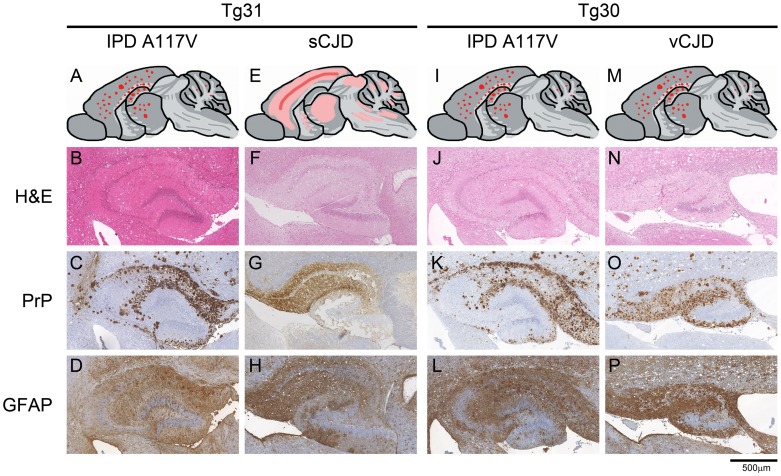
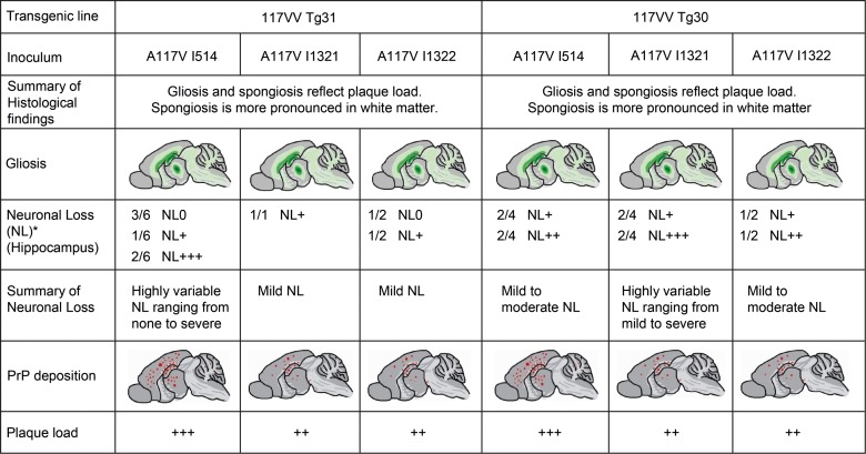
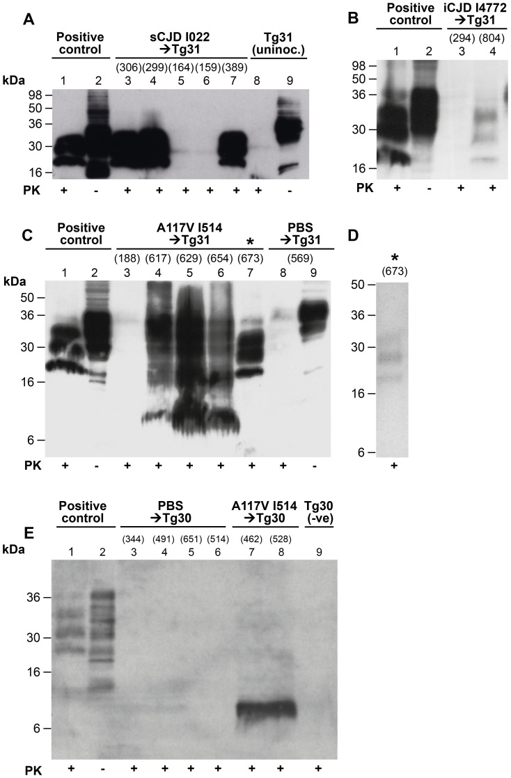
Prions are infectious agents causing fatal neurodegenerative diseases of humans and animals. In humans, these have sporadic, acquired and inherited aetiologies. The inherited prion diseases are caused by one of over 30 coding mutations in the human prion protein (PrP) gene (PRNP) and many of these generate infectious prions as evidenced by their experimental transmissibility by inoculation to laboratory animals. However, some, and in particular an extensively studied type of Gerstmann-Sträussler-Scheinker syndrome (GSS) caused by a PRNP A117V mutation, are thought not to generate infectious prions and instead constitute prion proteinopathies with a quite distinct pathogenetic mechanism. Multiple attempts to transmit A117V GSS have been unsuccessful and typical protease-resistant PrP (PrP(Sc)), pathognomonic of prion disease, is not detected in brain. Pathogenesis is instead attributed to production of an aberrant topological form of PrP, C-terminal transmembrane PrP ((Ctm)PrP). Barriers to transmission of prion strains from one species to another appear to relate to structural compatibility of PrP in host and inoculum and we have therefore produced transgenic mice expressing human 117V PrP. We found that brain tissue from GSS A117V patients did transmit disease to these mice and both the neuropathological features of prion disease and presence of PrP(Sc) was demonstrated in the brains of recipient transgenic mice. This PrP(Sc) rapidly degraded during laboratory analysis, suggesting that the difficulty in its detection in patients with GSS A117V could relate to post-mortem proteolysis. We conclude that GSS A117V is indeed a prion disease although the relative contributions of (Ctm)PrP and prion propagation in neurodegeneration and their pathogenetic interaction remains to be established.


Conflict of interest statement

JC and JW are shareholders and JC is a director of D-Gen Limited, an academic spin-out company working in the field of prion disease diagnosis, decontamination and therapeutics. D-Gen markets one of the routine antibodies (ICSM 35) used in this study. This does not alter our adherence to all PLoS Pathogens policies on sharing data and materials.

Figures

Figure 1
Figure 1. Neuropathological analysis of transgenic mouse brain.
Panels A, E, I and M, schematics showing regional distribution of abnormal PrP deposits. Note that these panels reflect the overall spatial distribution of neuropathology and are not meant to indicate precise representations of individual brains. Panels B, F, J and N, H&E staining demonstrating spongiform neurodegeneration in the hippocampal areas. Panels C, G, K and O, PrP immunohistochemistry using anti-PrP monoclonal antibody ICSM 35 demonstrates abnormal PrP immunoreactivity. Panels D, H, L and P, GFAP staining demonstrating gliosis in the hippocampal areas. A–D, IPD A117V prions inoculated to 117VV Tg31 mouse; E–H, sporadic CJD prions inoculated to 117VV Tg31 mouse showing distinctive diffuse synaptic PrP deposition characteristic of sCJD prions (panel G); I–L, IPD A117V prions inoculated to 117VV Tg30 mouse. M–P, vCJD prions inoculated to 117VV Tg30 mouse showing abundant non-florid PrP plaques (panel O). Scale bar = 500 µm for all panels except A, E, I and M.
Figure 2
Figure 2. Overview of histological findings in 117VV HuPrP transgenic mice challenged with A117V prion isolates.
The figures are schematic drawings reflecting the overall spatial distribution and intensity pattern of the gliosis or PrP deposition within the experimental groups. They are not meant to indicate precise representations of individual brains. * Definition of values for neuronal loss: NL0: No neuronal loss; NL+: Drop out of single neurones either focally or within the Ammon's horn (AH), leaving the AH continuity intact; NL++: Focal or regional drop out, interrupting the continuity of the AH and creating a small-medium gap (up to 1/3 of the length of the AH); NL+++: Neuronal drop out leaving gaps of more than 1/3 of the AH's length. Ratios represent the proportion of samples with the corresponding neuronal loss score.
Figure 3
Figure 3. Immunoblot analysis of abnormal PrP propagated in the brains of 117VV transgenic mice challenged with IPD A117V and classical CJD.
Mice were inoculated with classical CJD and GSS A117V brain. Immunoblots were analysed by enhanced chemiluminescence with monoclonal anti-PrP antibody ICSM 35. The numbers in parentheses above relevant lanes, represent the number of days each mouse survived post-inoculation. The provenance of each brain sample is designated above each lane. (A) Immunoblots of sporadic CJD-inoculated 117VV Tg31 mice (lanes 3–7) showing PrPSc resistant to harsh proteinase K (PK) digestion performed with 100 µg/ml PK at 37°C for 1 h (lanes 3, 4, and 7). Positive control was from a transgenic mouse expressing wild type HuPrP-129MV challenged with the same CJD inoculum (lanes 1and 2). An uninoculated 117VV Tg31 mouse brain is shown in lanes 8 and 9. (B) Brain homogenate of a 117VV Tg31 mouse inoculated with iatrogenic CJD prions that died without clinical disease at 804 days post-infection, showing weakly detectable PrPSc partially resistant to harsh PK digestion of 100 µg/ml for 1 hour at 37°C (lane 4) compared to the same control as in Figure 3A (lanes 1 and 2). Brain homogenate of a mouse killed relatively early at 294 days post-inoculation, shows no detectable PrPSc (lane 3). (C) Immunoblots of brains of five separate 117VV Tg31 mice all inoculated with the same GSS A117V patient brain homogenate showing the presence of PrPC, PrPSc and 8 kDa PrP fragment following harsh PK digestion at 100 µg/ml PK at 37°C for 1 hour (lanes 3–7). Under these conditions PBS-inoculated age-matched control 117VV Tg31 mouse brain shows only residual PrPC signal on long exposure (lane 8). Brain homogenate of a mouse culled relatively early at 188 days post-inoculation, compared to the group mean survival post-inoculation of >616 days, showed no detectable PrPSc (lane 3). One 117VV Tg31 mouse was clinically sick at 673 days post-infection, and its brain sample shows complete digestion of PrPC and the presence of classical PrPSc (lane 7, denoted by *) confirming adequacy of the PK digestion conditions. (D) Immunoblotting was repeated for all samples shown in lanes 4–7 of Figure 3C. These samples had undergone only one further freeze–thaw cycle before PK digestion. Compared with the readily detectable abnormal PrP signals seen in Figure 3C, only one sample (denoted by *) now showed the presence of classical PrPSc but at reduced signal strength, and only after using PK at a reduced concentration of 10 µg/ml. In other samples, only an 8 kDa PrP fragment could be variably detected but after using reduced PK concentrations (see Figure S2B lanes 3 and 4). (E) Immunoblot showing only the 8 kDa PrP fragment associated with A117V-challenged 117VV Tg30 mouse brains analysed with 50 µg/ml PK at 37°C for 1 hour (lanes 7 and 8), whereas PrP in brain homogenates of PBS-challenged Tg30 mice (lanes 3–6) and uninoculated age-matched Tg30 mouse brain (lane 9) is completely digested under the same conditions. Positive control in lanes 1 and 2 is brain homogenate of a transgenic mouse expressing wild type HuPrP (129MM Tg35) that was challenged with classical CJD.
Figure 4
Figure 4. Immunoblot analysis of abnormal PrP propagated in the brains of 117VV Tg30 transgenic mice challenged with vCJD prions.
All mice were inoculated with the same vCJD prion isolate. Immunoblots were analysed by enhanced chemiluminescence with monoclonal anti-PrP antibody ICSM 35. The provenance of the brain samples is designated above the lanes. (A) Lanes 3 and 4 show the predicted type 5 PrPSc bands seen when vCJD is propagated in transgenic mice expressing HuPrP with the codon 129VV genotype, compared with the detection of type 4 PrPSc in the brain of vCJD-inoculated 129MM Tg45 mouse (lanes 1 and 2). Type 5 shares the glycoform ratio of type 4 but differs in migrating more slowly on western blots because all 3 glycoform fragments of type 5 have higher apparent molecular masses than those of type 4. The lower signal intensity in lane 3 (100 µg/ml PK) compared to lane 4 (50 µg/ml PK) reflects PK-sensitivity of the vCJD-seeded 117V PrPSc. The 8 kDa PrP fragments can be seen associated with only PK-digested prion-infected 117V PrP-expressing mouse brain samples. These truncated 8 kDa fragments are absent from either vCJD infected Tg45 mice (lanes 1 and 2) or vCJD-infected 117VV Tg30 brain samples not digested with PK (lane 5).These data confirm that the 8 kDa human PrP fragment is a disease-associated PrP degradation product. (B) Variable PK resistance in brain of 117VV Tg30 mouse inoculated with vCJD. The 8 kDa PrP fragment is only seen in PK digested lanes 1–4, but is absent in the same sample undigested with PK (lane 5).

References

    1. Griffith JS (1967) Self replication and scrapie. Nature 215: 1043–1044. - PubMed
    1. Prusiner SB (1982) Novel proteinaceous infectious particles cause scrapie. Science 216: 136–144. - PubMed
    1. Collinge J, Clarke A (2007) A general model of prion strains and their pathogenicity. Science 318: 930–936. - PubMed
    1. Collinge J (2001) Prion diseases of humans and animals: their causes and molecular basis. Ann Rev Neurosci 24: 519–550. - PubMed
    1. Hill AF, Joiner S, Linehan J, Desbruslais M, Lantos PL, et al. (2000) Species barrier independent prion replication in apparently resistant species. Proc Natl Acad Sci USA 97: 10248–10253. - PMC - PubMed

Publication types