. 2013 Sep 25;8(9):e75248.
doi: 10.1371/journal.pone.0075248. eCollection 2013.

Ultrastructural abnormalities in CA1 hippocampus caused by deletion of the actin regulator WAVE-1

Affiliations

Ultrastructural abnormalities in CA1 hippocampus caused by deletion of the actin regulator WAVE-1

Diána Hazai et al. PLoS One. .

Abstract

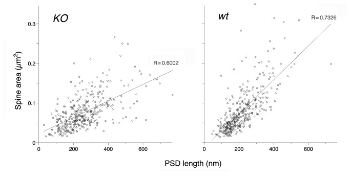
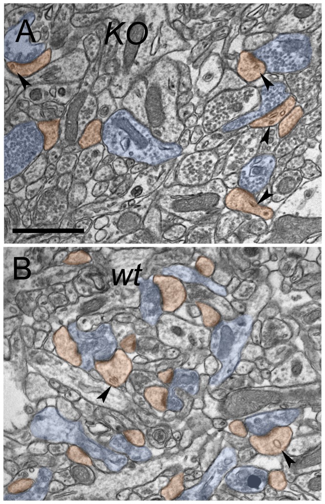
By conveying signals from the small GTPase family of proteins to the Arp2/3 complex, proteins of the WAVE family facilitate actin remodeling. The WAVE-1 isoform is expressed at high levels in brain, where it plays a role in normal synaptic processing, and is implicated in hippocampus-dependent memory retention. We used electron microscopy to determine whether synaptic structure is modified in the hippocampus of WAVE-1 knockout mice, focusing on the neuropil of CA1 stratum radiatum. Mice lacking WAVE-1 exhibited alterations in the morphology of both axon terminals and dendritic spines; the relationship between the synaptic partners was also modified. The abnormal synaptic morphology we observed suggests that signaling through WAVE-1 plays a critical role in establishing normal synaptic architecture in the rodent hippocampus.


Conflict of interest statement

Competing Interests: The authors have declared that no competing interests exist.

Figures

Figure 1
Figure 1. Overview of ultrastructural changes associated with deletion of WAVE-1.
Representative low-magnification electron micrographs of synaptic neuropil in stratum radiatum of CA1 hippocampus, showing postsynaptic spines (orange) and presynaptic boutons (blue) in material from KO (A) and wt mice (B). Scale bar: 5 µm.
Figure 2
Figure 2. WAVE-1 affects size of presynaptic terminals and organization of synaptic vesicles.
A. Cumulative plot shows the distribution of the mean diameter (defined as sqrt(area*4/π)) of terminals in KO (grey circles) and wt (black diamonds) CA1 KO terminals are generally larger than wt terminals. B. Electron micrographs (upper panel) and corresponding line drawings (lower panel) illustrate organization of synaptic vesicles within an axon terminal from a KO mouse (left), compared to wt control (right). Micrographs are from stratum radiatum of CA1 hippocampus. Synaptic vesicles are more numerous and lie farther from the active zone in KO mice, compared to wt. Scale bar: 200 nm. C. Quantitative analysis of the organization of vesicles in KO mice, versus wt controls. To combine data from terminals of different sizes, the distribution of vesicles was normalized (see inset): 0 corresponds to a vesicle lying directly at the presynaptic membrane, and 1 to a vesicle lying at the opposite non-synaptic membrane along an axis perpendicular to the synapse. Black circles (representing positions of KO vesicles in terminals) tend to lie farther from the active zone than white circles (representing positions of the wt vesicles). Vertical bars are standard errors (N = 3 animals for each genotype).
Figure 3
Figure 3. WAVE-1 affects relationship between PSD size and spine size.
Scatterplots show the area of spine profiles as a function of PSD length, in CA1 stratum radiatum of KO (left) and wt hippocampus (right). Linear regression analysis demonstrates a weaker correlation between spine area and PSD length in the KO (R = 0.60) than the wt material (R = 0.73).
Figure 4
Figure 4. Loss of WAVE-1 causes abnormalities in the internal structure of spines.
Representative electron micrographs of CA1 synaptic neuropil; coloring as in Figure 1. Spines from the KO material (A) were far more likely to contain endosomes (black arrowheads) than spines from the wt (B). Scale bar: 1 µm.
Figure 5
Figure 5. Abnormal spines in stratum radiatum of the WAVE-1 KO mouse.
A. Arrows point to spines with two PSDs. Spines are colored in orange, axon terminals in blue. B. 3D reconstruction of an axospinous synaptic contact from mutant hippocampus. The reconstructed CA1 apical dendritic segment (D) has a spine (Sp) oriented to show the synaptic surface. This spine has two distinct postsynaptic densities (red, arrows). The same axon (yellow) establishes separate synaptic contacts with both PSDs. Axon is yellow, spine is grey, PSD is red. Arrows point to synaptic surface between presynaptic active zone and postsynaptic density of the spine (Sp). Scale bars: 200 nm.

References

    1. Fortin DA, Srivastava T, Soderling TR (2012) Structural modulation of dendritic spines during synaptic plasticity. Neuroscientist 18: 326-341. doi:10.1177/1073858411407206. PubMed: 21670426. - DOI - PubMed
    1. Lippman J, Dunaevsky A (2005) Dendritic spine morphogenesis and plasticity. J Neurobiol 64: 47-57. doi:10.1002/neu.20149. PubMed: 15884005. - DOI - PubMed
    1. Nakagawa T, Engler JA, Sheng M (2004) The dynamic turnover and functional roles of alpha-actinin in dendritic spines. Neuropharmacology 47: 734-745. doi:10.1016/j.neuropharm.2004.07.022. PubMed: 15458845. - DOI - PubMed
    1. Matus A (2000) Actin-based plasticity in dendritic spines. Science 290: 754-758. doi:10.1126/science.290.5492.754. PubMed: 11052932. - DOI - PubMed
    1. Fifková E, Delay RJ (1982) Cytoplasmic actin in neuronal processes as a possible mediator of synaptic plasticity. J Cell Biol 95: 345-350. doi:10.1083/jcb.95.1.345. PubMed: 6890558. - DOI - PMC - PubMed

Publication types

Substances

LinkOut - more resources