. 2013 Sep 25;8(9):e75854.
doi: 10.1371/journal.pone.0075854. eCollection 2013.

Induction of cytopathogenicity in human glioblastoma cells by chikungunya virus

Affiliations

Induction of cytopathogenicity in human glioblastoma cells by chikungunya virus

Rachy Abraham et al. PLoS One. .

Abstract

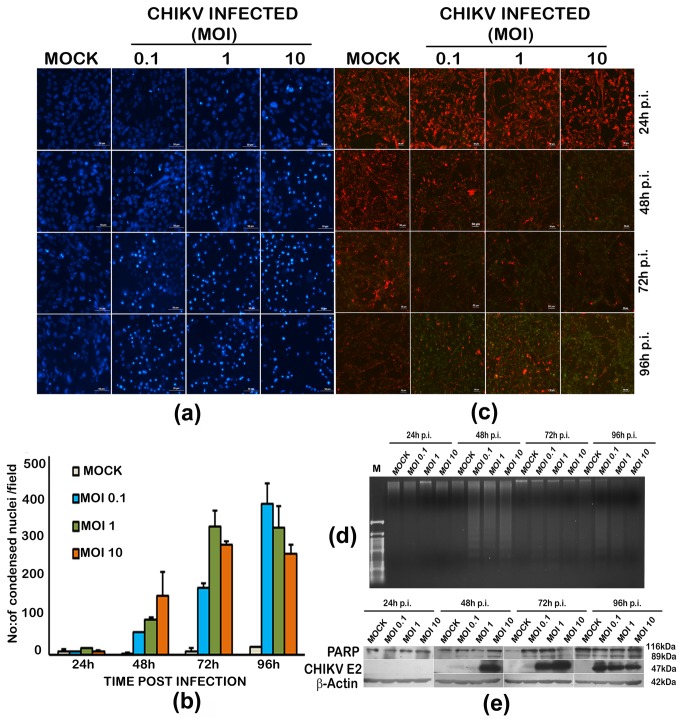
Chikungunya virus (CHIKV), an arthritogenic old-world alphavirus, has been implicated in the central nervous system (CNS) infection in infants and elderly patients. Astrocytes are the major immune cells of the brain parenchyma that mediate inflammation. In the present study we found that a local isolate of CHIKV infect and activate U-87 MG cells, a glioblastoma cell line of human astrocyte origin. The infection kinetics were similar in infected U-87 MG cells and the human embryo kidney (HEK293) cells as indicated by immunofluorescence and plaque assays, 24h post-infection (p.i.). In infected U-87 MG cells, apoptosis was detectable from 48h p.i. evidenced by DNA fragmentation, PARP cleavage, loss of mitochondrial membrane potential, nuclear condensation and visible cytopathic effects in a dose and time-dependent manner. XBP1 mRNA splicing and eIF2α phosphorylation studies indicated the occurrence of endoplasmic reticulum stress in infected cells. In U-87 MG cells stably expressing a green fluorescent protein-tagged light chain-3 (GFP-LC3) protein, CHIKV infection showed increased autophagy response. The infection led to an enhanced expression of the mRNA transcripts of the pro-inflammatory cytokines IL-1β, TNF-α, IL-6 and CXCL9 within 24h p.i. Significant up-regulation of the proteins of RIG-I like receptor (RLR) pathway, such as RIG-I and TRAF-6, was observed indicating the activation of the cytoplasmic-cellular innate immune response. The overall results show that the U-87 MG cell line is a potential in vitro model for in depth study of these molecular pathways in response to CHIKV infection. The responses in these cells of CNS origin, which are inherently defective in Type I interferon response, could be analogous to that occurring in infants and very old patients who also have a compromised interferon-response. The results also point to the intriguing possibility of using this virus for studies to develop oncolytic virus therapy approaches against glioblastoma, a highly aggressive malignancy.


Conflict of interest statement

Competing Interests: The authors have declared that no competing interests exist.

Figures

Figure 1
Figure 1. Infection of U-87 MG glioblastoma cells and HEK 293 cells with chikungunya virus.
Monolayer cultures of the cells were infected at different multiplicity of infection (MOI) and observed for different duration post-infection (p.i.). Uninfected cells were kept as control. (a) Bright field microscopic images of the infected HEK293 cells. (b) Immunofluorescence detection of CHIKV infection in HEK293 cells. CHIKV infected and control cells were fixed using 4% paraformaldehyde and subjected to immunofluorescence analysis as described in methods section. The virus infection was detected using an in-house anti-CHIKV E1 envelope protein rabbit polyclonal serum at 1:10 dilution. FITC conjugated anti-rabbit IgG was used as the secondary antibody. The presence of infection is indicated by the green fluorescence foci in the infected cells. A secondary antibody control did not show any background staining at the dilutions used in the experiment (not shown). (c) Bright field microscopic images showing the cytopathic changes in the infected U-87 MG cells. Changes such as rounding and intracytoplasmic granulation are visible at 72h p.i, but they are much more pronounced at 96h p.i. The control cells remain intact and healthy at 96h p.i. Scale bar represents 50µm. (d) Immunofluorescence detection of CHIKV infection in U-87 MG cells. (e) & (f) Kinetics of virus release in the culture supernatants from CHIKV infected HEK293 cells (e) and U-87 MG cells (f). Culture supernatants were collected at various time points post-infection and the virus plaque forming units were detected separately in confluent monolayer cultures of Vero cells as described in the methods. The values indicate the average of the number of plaques from three independent experiments, each in turn calculated from three dilutions of the sample giving <100 plaques, and represented as log10 values.
Figure 2
Figure 2. Induction of apoptosis by chikungunya virus in U-87 MG cells.
(a) Evidence of nuclear condensation in infected cells upon Hoechst staining. Monolayer cultures of the cells were infected at different multiplicity of infection (MOI) and observed for different duration post-infection (p.i.). Uninfected cells were kept as control. Cells were stained with nuclear staining dye Hoechst 33342 as described in methods section and viewed under a fluorescent microscope. Scale bar represent 50µm. (b) Quantitative estimation of the condensed nuclei in infected cells. Each value is an average of four fields from three independent experiments. (c) Mitochondrial membrane potential loss in infected cells. Cells were stained with JC1 dye and the membrane potential loss is seen as loss of red fluorescence and increase in green fluorescence pronounced at 96h p.i. (d) DNA fragmentation analysis by Agarose gel electrophoresis. Total genomic DNA (1µg) isolated from CHIKV infected and uninfected cells were subjected to electrophoresis in a 0.8% agrose gel and visualized by ethidium bromide staining in a UV transilluminator. DNA fragmentation indicating apoptosis is visible in infected cells from 48h onwards. (e) Western blot of infected and uninfected cells. PARP-cleavage evidenced by presence of an 89kDa cleaved protein in infected cells. Detection of envelope protein expression using anti-CHIKV E2 antibody was used for confirming presence of the virus. β-actin expression serves as the loading control.
Figure 3
Figure 3. Induction of autophagy and endoplasmic reticulum stress in chikungunya virus infected U-87 MG cells.
(a) Punctae formation indicating the autophagosomes. U-87 MG cells stably transfected with a GFP-tagged light chain 3 (LC3) expression-construct, and cells transiently transfected with GFP expressing plasmid alone were infected with CHIKV, and observed 24h post-infection. Distinct punctae formation by LC3 recruitment to autophagosomes is visible in the infected cells expressing GFP-LC3. (b) Quantitation of the autophagy response. The average number of puncta/field in infected and uninfected cells is given. Values are Mean ±SD from four different microscopic fields and two independent experiments. ‘**’ indicates significance level p<0.005 (c) XBP-1 mRNA splicing indicating ER stress in infected cells. Total RNA was isolated from infected and uninfected cells at different time points and subjected to reverse-transcription PCR and agarose gel electrophoresis as described in the methods. The splice variant of XBP-1 (263bp) along with the original mRNA (289bp) is detectable in CHIKV infected cells from as early as 12h post-infection, but is more pronounced at 72h post infection in cells infected at an MOI of 10.Amplification of β-actin mRNA serves as the loading control. (d) eIF2α phosphorylation in CHIKV infected U-87 MG cells. Total cell lysate from the infected and uninfected cells was isolated 24h, 48 and 72h p.i. 50µg of total protein was subjected to SDS-PAGE and western blot as described in the methods using anti- eIF2α and anti-phopho-eIF2α antibodies. The band intensities of phopho-eIF2α and eIF2α expression, determined from densitometric analysis, were initially normalised independently to that of corresponding β-Actin, and the ratio of phopho- eIF2α to eIF2α expression was calculated. The fold change in expression was calculated relative to the protein expression in uninfected controls at the respective time points. Values are Mean ± SD from three independent experiments.
Figure 4
Figure 4. Modulation of the transcripts of pro-inflammatory cytokines in CHIKV infected U-87 MG cells.
Total RNA from the infected and uninfected cells was isolated 24h p.i and was subjected to semi-quantitative RT-PCR as described in the methods. The PCR band intensities of the cytokine cDNA amplicons, determined from densitometric analysis after agarose-gel electrophoresis, were initially normalised to that of corresponding β-Actin amplicon, and the fold change in expression was calculated relative to the gene expression in uninfected controls at the respective time-points. Values are Mean ± SD from three independent biological experiments each done in duplicate. ‘*’ indicates statistically significant (p<0.05) modulation with respect to the basal level (0h) gene expression.
Figure 5
Figure 5. Modulation of the innate immune response sensors in CHIKV infected U-87 MG cells.
A: Semi-quantitative RT-PCR. Total RNA from the infected and uninfected cells was isolated 24h p.i and was subjected to semi-quantitative RT-PCR as described in the methods. The PCR band intensities of the target cDNA amplicons, determined from densitometric analysis after agarose-gel electrophoresis, were initially normalised to that of corresponding β-Actin amplicon, and the fold change in expression was calculated relative to the gene expression in uninfected controls at the respective time-points. Values are Mean ± SD from three independent biological experiments each done in duplicate. ‘*’ indicates significant modulation with respect to the basal level (0h) gene expression. B. Western blot and quantitation by densitometric analysis. Total cell lysate from the infected and uninfected cells was isolated 24h p.i and 50-100µg of total protein was subjected to SDS-PAGE and western blot as described in the methods. The band intensities of the targets, determined from densitometric analysis, were initially normalised to that of corresponding β-Actin, and the fold change in expression was calculated relative to the protein expression in uninfected controls at 24h p.i. Values are Mean ± SD from three independent experiments. ‘*’ indicates statistically significant (p<0.05) modulation with respect to the expression in the uninfected cells.

References

    1. Das T, Jaffar-Bandjee MC, Hoarau JJ, Krejbich Trotot P, Denizot M et al. (2010) Chikungunya fever: CNS infection and pathologies of a re-emerging arbovirus. Prog Neurobiol 91: 121-129. doi:10.1016/j.pneurobio.2009.12.006. PubMed: 20026374. - DOI - PubMed
    1. Arpino C, Curatolo P, Rezza G (2009) Chikungunya and the nervous system: what we do and do not know. Rev Med Virol 19: 121-129. doi:10.1002/rmv.606. PubMed: 19274635. - DOI - PubMed
    1. Lewthwaite P, Vasanthapuram R, Osborne JC, Begum A, Plank JL et al. (2009) Chikungunya virus and central nervous system infections in children, India. Emerg Infect Dis 15: 329-331. doi:10.3201/eid1502.080902. PubMed: 19193287. - DOI - PMC - PubMed
    1. Chandak NH, Kashyap RS, Kabra D, Karandikar P, Saha SS et al. (2009) Neurological complications of Chikungunya virus infection. Neurol India 57: 177-180. doi:10.4103/0028-3886.51289. PubMed: 19439849. - DOI - PubMed
    1. Griffin DE (2010) Emergence and re-emergence of viral diseases of the central nervous system. Prog Neurobiol 91: 95-101. doi:10.1016/j.pneurobio.2009.12.003. PubMed: 20004230. - DOI - PMC - PubMed