. 2013 Sep 25;8(9):e76038.
doi: 10.1371/journal.pone.0076038. eCollection 2013.

Anti-HIV activity of human defensin 5 in primary CD4+ T cells under serum-deprived conditions is a consequence of defensin-mediated cytotoxicity

Affiliations

Anti-HIV activity of human defensin 5 in primary CD4+ T cells under serum-deprived conditions is a consequence of defensin-mediated cytotoxicity

Jian Ding et al. PLoS One. .

Abstract

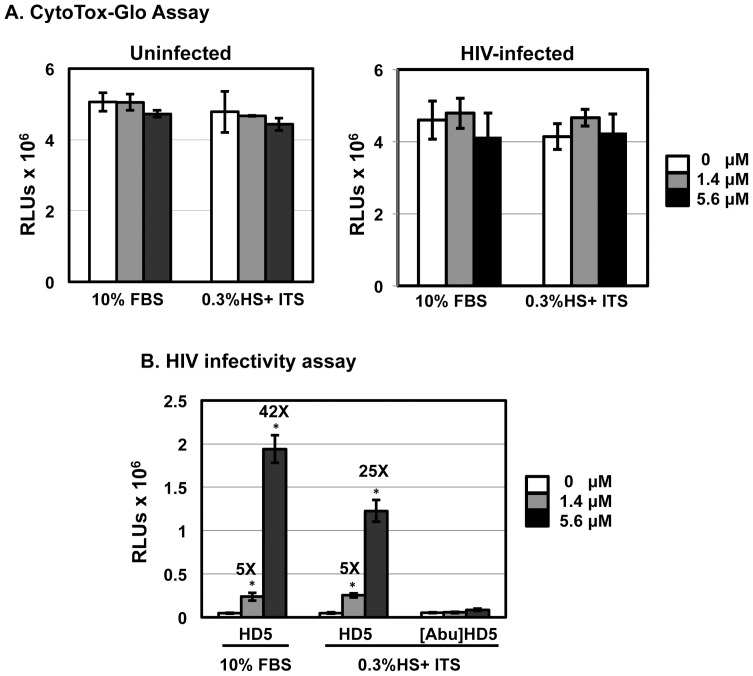
Background: We have previously shown that human defensin 5 (HD5) promotes HIV infectivity in both primary CD4+ T cells and HeLa cells expressing CD4 and CCR5. HD5 is induced in response to sexually transmitted infections (STIs) such as Chlamydia trachomatis and Neisseria gonorrhoeae, suggesting it plays a role in STI-mediated enhancement of HIV transmission. In contrast to our findings, a recent study reports that HD5 has an anti-HIV effect in primary CD4+ T cells under serum-deprived conditions. To resolve these apparently contradictory observations, we investigated experimental parameters that might contribute to contrasting effects of HD5.

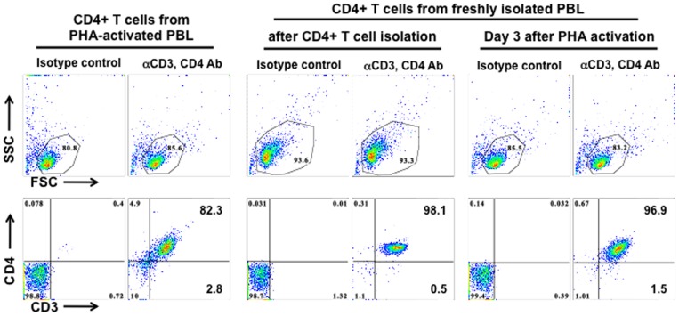
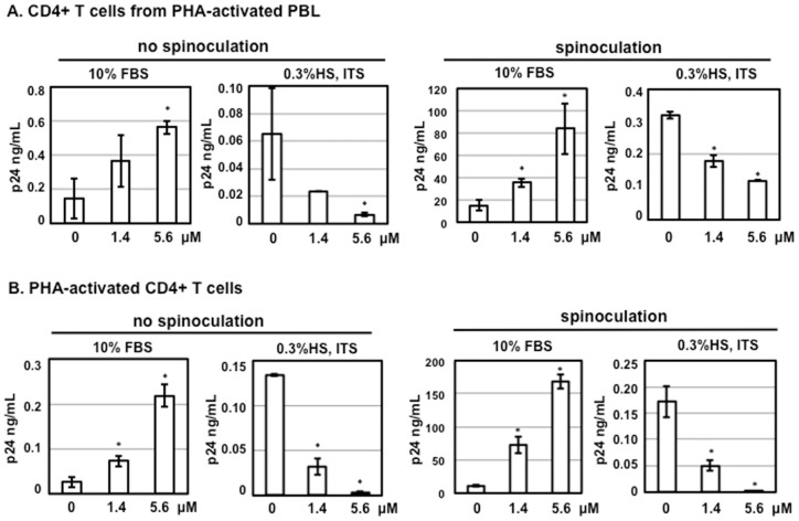
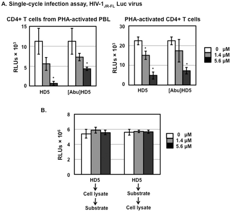
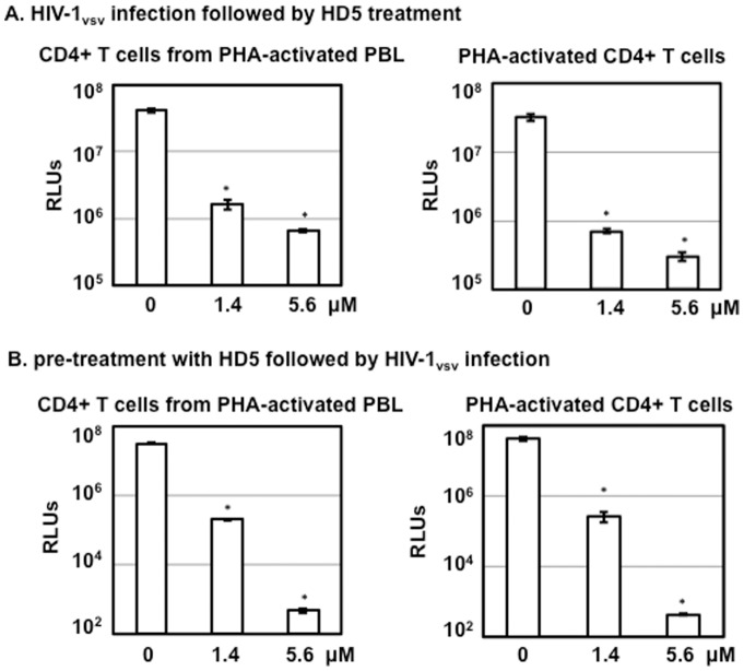
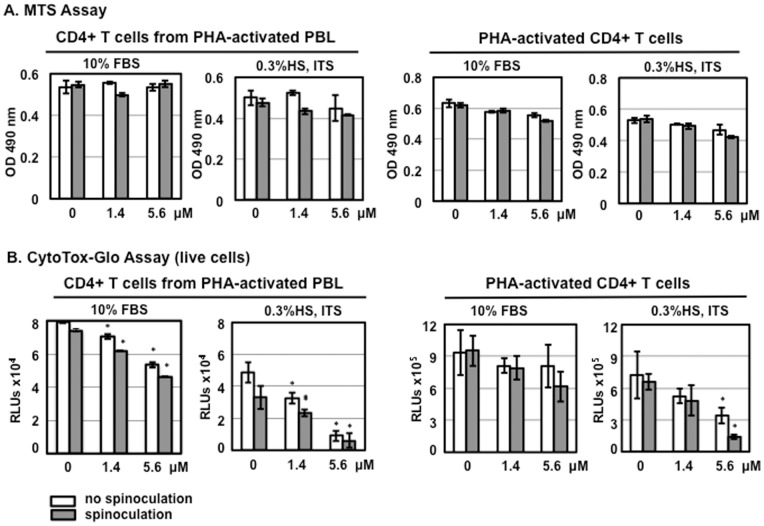
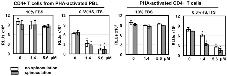
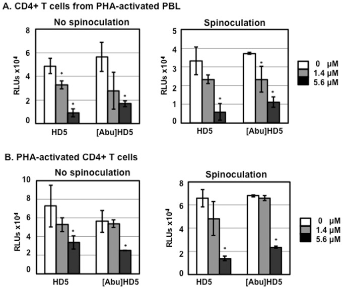
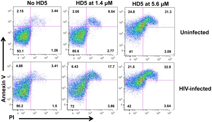
Results: Serum-deprived culture conditions were associated with anti-HIV activity. In contrast to the dependence of the HIV enhancing effect on HD5 structure, the anti-HIV activity in serum-deprived primary CD4+ T cells was independent of HD5 structure as the linear peptide [Abu] HD5 exhibited similar anti-HIV activity. Under serum deprived conditions, HD5 blocked CD4-receptor-independent HIV-1vsv infection before or after viral entry. We found that HD5 and its linear form induced significant cell death in primary CD4+ T cells under serum-deprived culture conditions. HD5-mediated apoptosis was observed as early as 2 h after addition of defensins to serum-deprived primary CD4+ T cells. In contrast to primary CD4+ T cells, HD5 did not induce cytotoxicity and promote HIV infectivity of HeLa-CD4-CCR5 cells under serum-deprived conditions.

Conclusions: These results indicate that under serum-deprived culture conditions HD5 is toxic for primary CD4+ T cells, warranting caution in data interpretation.


Conflict of interest statement

Competing Interests: The authors have declared that no competing interests exist.

Figures

Figure 1
Figure 1. CD4+ T cells from PHA-activated PBLs are less pure than cells prepared from freshly isolated PBLs.
CD4+ T cells isolated from PHA-activated PBLs (left panel), CD4+ T cells right after isolation from fresh PBLs (middle panel), or CD4+ T cells isolated from fresh PBLs followed by PHA activation for 3 days (right panel) were stained with PE-conjugated mouse anti-CD3 Ab and PerCP-conjugated mouse anti-CD4 Ab. Gated live cells in the scatter plot are shown in the upper panels. The results shown are representative of 3 tested donors, which are summarized in Table 1.
Figure 2
Figure 2. Serum deprivation contributes to anti-HIV activity of HD5 in primary CD4+ T cells.
HIV-1 primary isolate 20635–4 (R5 virus, clade C) was incubated with HD5 at different concentrations for 1 h at 37°C. CD4+ T cells from PHA-activated PBLs (A) and PHA-activated CD4+ T cells (B) were incubated with virus-defensin mixture for 2 h at 37°C (no spinoculation) or 1.5 h at 1250×g spinning (spinoculation). After washing off unbound virus, cells were cultured in RPMI containing 10% FBS and IL-2 or 0.3% human AB serum, ITS supplement, and IL-2. HD5 was added back to cell cultures in the presence of IL-2. The level of p24 protein in culture media was measured by p24 ELISA. Data presented are the average ± standard deviation of 3 replicates. Similar results were observed in 3 independent experiments from different donors; *P<0.05, defensin-treated samples vs non-treated controls.
Figure 3
Figure 3. HD5 and [Abu]HD5 inhibit HIV infection in serum-deprived primary CD4+ T cells.
(A). Serum-free HIV-1JR-FL Env-pseudotyped reporter viruses were incubated with HD5 or its linear peptide, [Abu]HD5 for 1 h at 37°C before addition to CD4+ T cells by spinoculation. After washing off unbound virus, cells were cultured under serum-deprived condition in the presence of defensins. HIV infection was determined by measuring luciferase activity at day 3 after infection. Data presented are the average ± standard deviation of 3 replicates; *P<0.05, defensin-treated samples vs non-treated controls. Similar results were observed in 2 independent experiments. (B) To determine whether HD5 interfered with luciferase activity in vitro, 50 µl of cell lysate from HeLa-CD4-CCR5 cells or 100 µl of luciferase substrate was incubated with HD5 at different concentrations for 60 min on ice. Then, 100 µl of luciferase assay substrate or 50 µl of cell lysate was added to the mixture, respectively, and luminescence was measured.
Figure 4
Figure 4. HD5-mediated inhibition of HIV in serum-deprived primary CD4+ T cells occurs independently of HIV receptors.
(A) CD4+ T cells from different preparation methods were infected with pseudotyped HIV-1VSV-G luciferase reporter viruses by spinoculation. Infected cells were cultured under serum-deprived conditions. HIV infection was determined by measuring luciferase activity on day 3 after infection. (B) CD4+ T cells under serum-deprived conditions were treated with HD5 at different concentrations for 1 h at 37°C followed by exposure to serum-free pseudotyped HIV-1VSV-G luciferase reporter viruses by spinoculation in the presence of HD5. Cells were then cultured in RPMI containing 0.3% human AB serum, ITS supplement, IL-2, and defensins for 3 days followed by measurement of luciferase activity. Data presented are the average ± standard deviation of 3 replicates and represent 2 independent experiments. *P<0.05, defensin-treated samples vs non-treated controls.
Figure 5
Figure 5. HD5 induces cytotoxicity in primary CD4+ T cells under serum-deprived conditions.
CD4+ T cells (1×10⁴ cells per sample) were treated with HD5 at different concentrations with or without spinoculation. Cells were then cultured in RPMI-1640 containing10% FBS and IL-2 or RPMI-1640 containing 0.3% human AB serum, 1× ITS supplement, and IL-2. HD5 was added back. After overnight culture, cell proliferation was measured by MTS assay (A); cytotoxicity was measured by CytoTox-Glo cytotoxicity kit (B). Data presented are the average ± standard deviation of 3 replicates; *P<0.05, defensin-treated samples vs non-treated controls. Similar results were observed in 3 independent experiments.
Figure 6
Figure 6. HD5 induces cytotoxicity in HIV-exposed primary CD4+ T cells under serum-deprived conditions.
HIV-1JR-FL was incubated with HD5 at different concentrations at 37°C for 1 h. CD4+ T cells were incubated with defensin-virus mixture with or without spinoculation as described in Fig. 2. Cells were washed and then cultured with the original concentration of HD5 in either RPMI-1640 containing10% FBS and IL-2 or RPMI-1640 containing 0.3% human AB serum, 1× ITS supplement, and IL-2. After overnight culture, cytotoxicity was measured by CytoTox-Glo cytotoxicity kit (Promega). Data presented are the average ± standard deviation of 3 replicates; *P<0.05, defensin-treated samples vs non-treated controls. Similar results were observed in 2 independent experiments.
Figure 7
Figure 7. HD5 and linear peptide [Abu]HD5 induced cytotoxicity in primary CD4+ T cells under serum-deprived conditions.
CD4+ T cells from PHA-activated PBLs (A) or PHA-activated CD4+ T cells (B) were exposed to HD5 or [Abu]HD5 at indicated concentrations with or without spinoculation. Cells were then cultured for 24 h under serum-deprived conditions in the presence of IL-2 and defensins. Cytotoxicity was measured by CytoTox-Glo cytotoxicity kit. Data presented are the average ± standard deviation of 3 replicates and represent 3 independent experiments. *P <0.05, defensin-treated samples vs non-treated controls.
Figure 8
Figure 8. HD5 induces apoptosis in PHA-activated CD4+ T cells and HIV-infected CD4+ T cells under serum-deprived conditions.
PHA-activated CD4+ T cells under serum-deprived conditions were treated with HD5 at different concentrations for 24 h before staining with Annexin V and PI. Activated CD4+ T cells were also exposed to defensin-treated HIV-1JR-FL for 2 h, washed and then treated with HD5. Apoptosis was determined by FACS analysis. Similar results were obtained using cells from two independent donors.
Figure 9
Figure 9. Anti-HIV activity and cytotoxicity of HD5 are not found in HeLa-CD4-CCR5 cells under serum-deprived condition.
(A) HeLa-CD4-CCR5 cells were exposed to HD5 at indicated concentrations in serum-free RPMI-1640 medium for 2 h at 37°C. The effect of HD5 on HIV-exposed HeLa-CD4-CCR5 cells was also prepared as described in Fig. 6 as a comparison. The cells were then cultured for 24 h in complete medium (10%FBS) or under serum-deprived conditions. Cytotoxicity was measured by CytoTox-Glo cytotoxicity kit. (B) Serum-free HIV-1JR-FL Env-pseudotyped reporter viruses were incubated with HD5 or [Abu]HD5 for 1 h at 37°C before exposure to HeLa-CD4-CCR5 cells for 2 h at 37°C. Cells were cultured in complete media or under serum-deprived conditions. Data presented are the average ± standard deviation of 3 replicates and represent 2 independent experiments. *P<0.05, defensin-treated samples vs non-treated controls.

References

    1. Ganz T (2003) Defensins: antimicrobial peptides of innate immunity. Nat Rev Immunol 3: 710–720. - PubMed
    1. Salzman NH (2010) Paneth cell defensins and the regulation of the microbiome: detente at mucosal surfaces. Gut microbes 1: 401–406. - PMC - PubMed
    1. Lehrer RI, Lu W (2012) alpha-Defensins in human innate immunity. Immunological reviews 245: 84–112. - PubMed
    1. Daher KA, Selsted ME, Lehrer RI (1986) Direct inactivation of viruses by human granulocyte defensins. J Virol 60: 1068–1074. - PMC - PubMed
    1. Ding J, Chou YY, Chang TL (2009) Defensins in viral infections. Journal of innate immunity 1: 413–420. - PMC - PubMed

Publication types