

Multifunctional nanorods serving as nanobridges to modulate T cell-mediated immunity
- PMID: 24088178
- PMCID: PMC3972813
- DOI: 10.1021/nn403275p
Multifunctional nanorods serving as nanobridges to modulate T cell-mediated immunity
Abstract
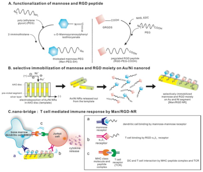
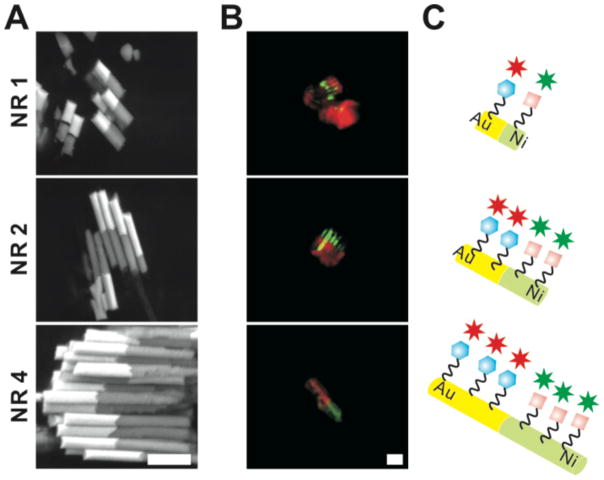
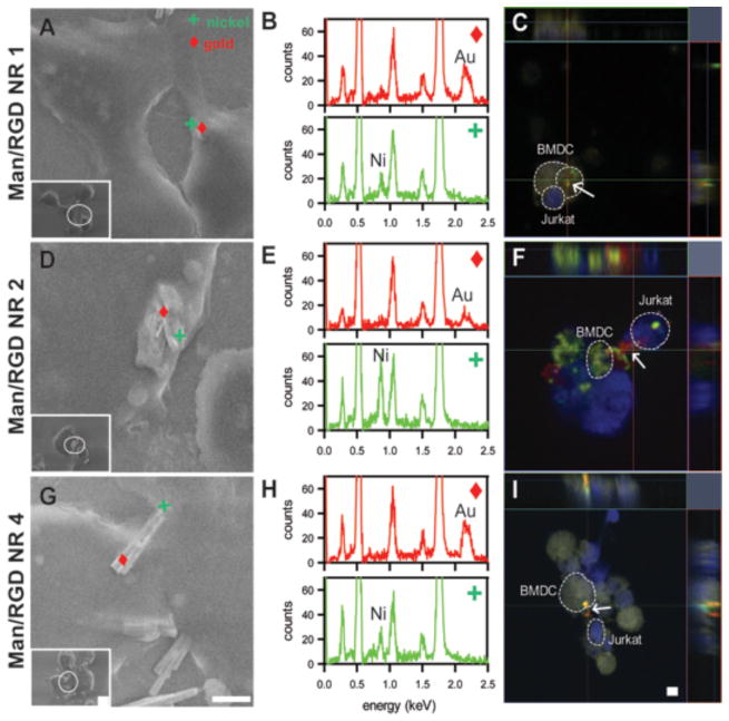
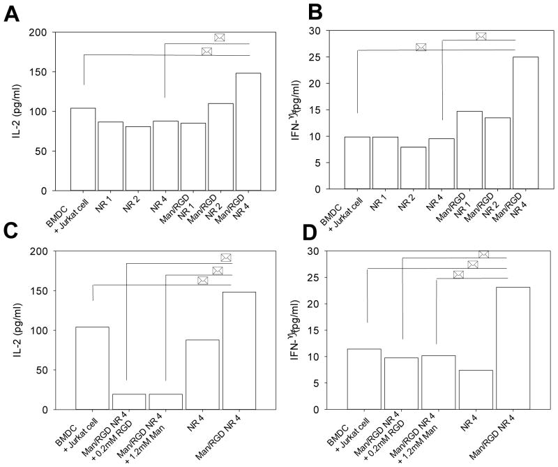
Electrodeposited nanorods serving as multivalent bridges were fabricated and surface-decorated with ligands for immune cells. Gold and nickel solutions were sequentially electrodeposited on nanoporous anodized disc templates and the template was dissolved to retrieve bisegmented nanorods with different lengths. Gold and nickel segmented nanorods were surface-immobilized with mannose and RGD peptides to prepare immune-cell recruiting nanorods. Surface-functionalization of nanorods were confirmed by fluorescence-labeling of each ligands and confocal microscopy. Dendritic cells and T cells were co-incubated with the surface-functionalized nanorods, and the proximity between the nanorods and the immune cells was visualized by variable pressure scanning electron microscopy and confocal microscopy. The long nanorods were associated with the immune cells, whereas the shorter nanorods were rather endocytosed by cells, suggesting a feasibility of the longer nanorods as bridging for the cells. Cytokine releases from the immune cells were monitored by cultivating lipopolysaccharide-activated dendritic cells with T cells. Interleukine-2 and interferon-γ release profiles showed a strong correlation with the length of the nanorod, where the 4 μm nanorods induced the highest levels of cytokine release compared to 1 or 2 μm nanorods. Thus, we concluded that the proximity of the immune cells increased by bridging the immune cells with the nanobridging system, which subsequently increased cytokine release by facilitating the antigen presentation process.
Figures
Similar articles
-
Applications, Surface Modification and Functionalization of Nickel Nanorods.Materials (Basel). 2017 Dec 28;11(1):45. doi: 10.3390/ma11010045. Materials (Basel). 2017. PMID: 29283415 Free PMC article. Review.
-
Photothermal reshaping of gold nanorods depends on the passivating layers of the nanorod surfaces.Langmuir. 2008 Oct 21;24(20):12026-31. doi: 10.1021/la800811j. Epub 2008 Aug 30. Langmuir. 2008. PMID: 18759472
-
RGD peptide immobilized on TiO2 nanotubes for increased bone marrow stromal cells adhesion and osteogenic gene expression.J Mater Sci Mater Med. 2012 Feb;23(2):527-36. doi: 10.1007/s10856-011-4479-0. Epub 2011 Dec 6. J Mater Sci Mater Med. 2012. PMID: 22143905
-
Segmented Pt/Ru, Pt/Ni, and Pt/RuNi nanorods as model bifunctional catalysts for methanol oxidation.Small. 2006 Jan;2(1):121-8. doi: 10.1002/smll.200500253. Small. 2006. PMID: 17193567
-
Interactions of Bacterial Lipopolysaccharides with Gold Nanorod Surfaces Investigated by Refractometric Sensing.ACS Appl Mater Interfaces. 2015 Nov 11;7(44):24915-25. doi: 10.1021/acsami.5b08440. Epub 2015 Nov 2. ACS Appl Mater Interfaces. 2015. PMID: 26488238
Cited by
-
Preparation of pyrenyl-based multifunctional nanocomposites for biomedical applications.Nat Protoc. 2016 Feb;11(2):236-51. doi: 10.1038/nprot.2015.135. Epub 2016 Jan 7. Nat Protoc. 2016. PMID: 26741408
-
Applications, Surface Modification and Functionalization of Nickel Nanorods.Materials (Basel). 2017 Dec 28;11(1):45. doi: 10.3390/ma11010045. Materials (Basel). 2017. PMID: 29283415 Free PMC article. Review.
-
Biofunctional Janus particles promote phagocytosis of tumor cells by macrophages.Chem Sci. 2020 May 5;11(20):5323-5327. doi: 10.1039/d0sc01146k. Chem Sci. 2020. PMID: 34122990 Free PMC article.
-
An Overview of the Use of Nanoparticles in Vaccine Development.Nanomaterials (Basel). 2023 Jun 9;13(12):1828. doi: 10.3390/nano13121828. Nanomaterials (Basel). 2023. PMID: 37368258 Free PMC article. Review.
-
Insights into the toxicological effects of nanomaterials on atherosclerosis: mechanisms involved and influence factors.J Nanobiotechnology. 2023 Apr 28;21(1):140. doi: 10.1186/s12951-023-01899-y. J Nanobiotechnology. 2023. PMID: 37118804 Free PMC article. Review.
References
-
- Kalishwaralal K, Banumathi E, Ram Kumar Pandian S, Deepak V, Muniyandi J, Eom SH, Gurunathan S. Silver Nanoparticles Inhibit Vegf Induced Cell Proliferation and Migration in Bovine Retinal Endothelial Cells. Colloids Surf B Biointerfaces. 2009;73:51–7. - PubMed
-
- Krippner-Heidenreich A, Tubing F, Bryde S, Willi S, Zimmermann G, Scheurich P. Control of Receptor-Induced Signaling Complex Formation by the Kinetics of Ligand/Receptor Interaction. J Biol Chem. 2002;277:44155–63. - PubMed
Publication types
MeSH terms
Substances
Grants and funding
LinkOut - more resources
Full Text Sources
Other Literature Sources
