. 2013 Oct 2:13:17.
doi: 10.1186/1472-6807-13-17.

Short tandem repeats in the inhibitory domain of the mineralocorticoid receptor: prediction of a β-solenoid structure

Affiliations

Short tandem repeats in the inhibitory domain of the mineralocorticoid receptor: prediction of a β-solenoid structure

Metaxia Vlassi et al. BMC Struct Biol. .

Abstract

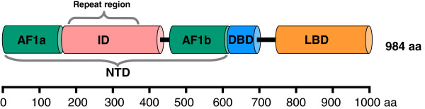
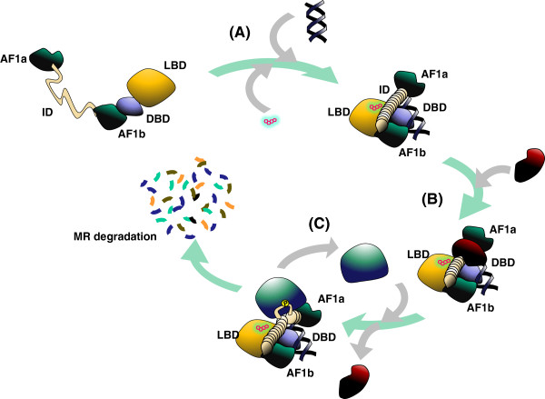
Background: The human mineralocorticoid receptor (MR) is one of the main components of the renin-angiotensin-aldosterone system (RAAS), the system that regulates the body exchange of water and sodium. The evolutionary origins of this protein predate those of renin and the RAAS; accordingly it has other roles, which are being characterized. The MR has two trans-activating ligand independent domains and one inhibitory domain (ID), which modulates the activity of the former. The structure of the ID is currently unknown.

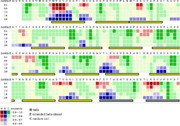
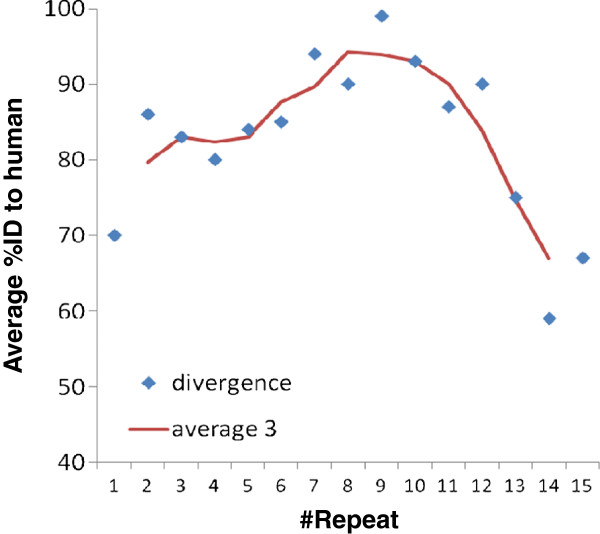
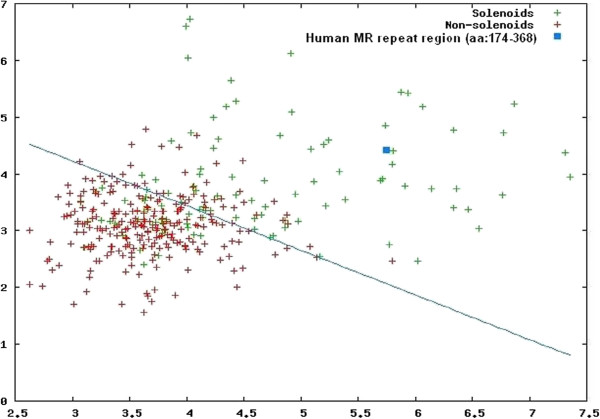
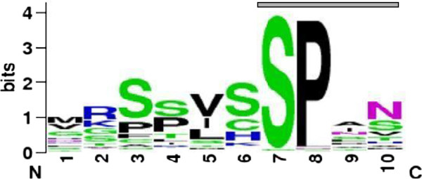
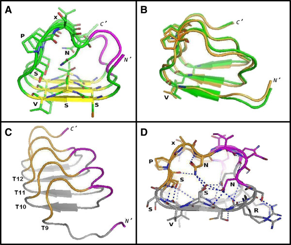
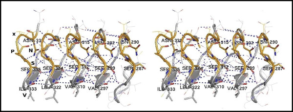
Results: Here we report that the ID contains at least 15 tandem repeats of around 10 amino acids, which we computationally characterize in the human MR and in selected orthologs. This ensemble of repeats seems to have emerged around 450 million years ago, after the divergence of the MR from its close homolog, the glucocorticoid receptor, which does not possess the repeats. The region would have quickly expanded by successive duplication of the repeats stabilizing at its length in human MR shortly after divergence of tetrapoda from bony fishes 400 million years ago. Structural predictions, in combination with molecular dynamics simulations suggest that the repeat ensemble forms a β-solenoid, namely a β-helical fold with a polar core, stabilized by hydrogen-bonded ladders of polar residues. Our 3D-model, in conjunction with previous experimental data, implies a role of the β-helical fold as a scaffold for multiple intra-and inter-molecular interactions and that these interactions are modulated via phosphorylation-dependent conformational changes.

Conclusions: We, thus, propose that the structure of the repeat ensemble plays an important role in the coordination and sequential interactions of various MR partners and therefore in the functionality and specificity of MR.


Figures

Figure 1
Figure 1
Domain structure of the human MR. The N-terminal domain (NTD) is composed of two activatory domains (AF1a and AF1b) and the inhibitory domain (ID). This is followed by the DNA binding domain (DBD) and the ligand (sterol) binding domain (LBD). We found a region with tandem repeats that covers most of the ID starting at its N-terminal. Other isoforms of the human MR have been defined but their splicing does not affect the tandem repeat region [4]. Isoform 1 is displayed (which is the longest one).
Figure 2
Figure 2
Multiple sequence alignment of the tandem repeat region. The positions of tandem repeats of length 10 aa are indicated with boxes in a multiple sequence alignment of the region with the repeats from the human MR with selected orthologs from tetrapoda and two bony fishes (Rutilus and zebrafish, Danio rerio) (see Methods for details). For some repeats, alignment between the tetrapoda and the fish repeat might not reflect a direct evolutionary relation but the similar amino acid properties of the repeats; this is indicated with separate boxes for fish repeats. Fish repeats F1 and F9 (red labels) have 6/10 identical positions, suggesting a recent event of repeat duplication in bony fishes. Matches to a regular expression used to identify the repeats ([GIKLMNRSTV] [ACEGNKPRSTV] [ACFGLMNPS] [AIKLPRSTV] [ADGILMPRSTV] [ACGHKRS] S P [AGHILMNPRSTV] [AGHIMNSTV]) are marked in yellow (see Methods for details). Tetrapoda repeats are labeled from #T1 to #T15. Fish repeats are labeled from #F1 to #F11. The region starts at the N-terminal of the ID. The last two repeats were identified with a complementary analysis (See text and Figure 3). The sequence identifiers indicate the Entrez Protein identifier and the species name for human (Homo sapiens), common marmoset (Callithrix jacchus), gray short-tailed opossum (Monodelphis domestica), dog (Canis lupus familiaris), panda (Ailuropoda melanoleuca), rat (Rattus norvegicus), mouse (Mus musculus), African clawed frog (Xenopus laevis), Carolina anole (a lizard, Anolis carolinensis), Zebra Finch (a bird, Taeniopygia guttata), chicken (Gallus gallus), the common roach (a fish, Rutilus Rutilus), and the zebrafish (Danio rerio). Known phosphorylation sites [8,9] are indicated by an asterisk (see text for details).
Figure 3
Figure 3
Contact-dependent secondary structure prediction of the repeat region in human MR. The CSSP profile [13] of the human MR tandem repeats (165-364 aa) using tertiary contacts (TC) in the range of 0.4 to 2.0 is shown. Predictions for α-helix, β-strand and random coil are colored red, blue and green, respectively, with variable propensity values, as indicated at the bottom of the figure. The position of predicted repeats is indicated under the alignment with boxed bars. Short β-strands are predicted upstream the conserved Ser-Pro motif of each repeat. This analysis confirmed the repeats identified in the pattern analysis (Figure 2) and suggested two additional (very divergent in sequence) repeats, marked with yellow and gray bars, respectively.
Figure 4
Figure 4
Similarity between tetrapoda repeats. The average percentage of identity to each human repeat (T1 to T15) from the corresponding repeats of the other tetrapoda included in the MSA shown in Figure 2 is plotted versus the position of the repeat. Higher values are observed for the middle repeats. The trend line (red) is an average of three consecutive repeats. The plot suggests higher conservation of the middle repeats.
Figure 5
Figure 5
Output of the REPETITA method. Scatter plot of optimal θ-ratio versus Zmax for the training sets and the human MR repeat region (aa: 174-368). These parameters reflect the existence of a periodic signal in several amino acid properties along the sequence and the largest spectral amplitude of those, respectively. Red and green crosses correspond to solenoid and non-solenoid proteins, respectively. The result corresponding to the human MR repeats is shown as a blue square.
Figure 6
Figure 6
Sequence logo representation of the MR repeat sequences. Font color indicates hydrophobic (black), basic (blue), polar (green) and asparagine (magenta) residues. The gray bar indicates the consensus sequence motif, SPxN. The figure was generated with WebLogo [22].
Figure 7
Figure 7
3D-models of consecutive MR repeats. (A) Initial model of three consecutive human MR repeats (T11 to T13, aa: 306-339). β-strands, turns and inter-repeat regions are colored in yellow, green and magenta, respectively. For clarity, only the side chains of residues discussed in the text are shown (stick models). The consensus sequence motifs, SSV and SPxN, are indicated. (B) Models resulting from the MD simulations. The dominant cluster of the last 50 ns of the REMD replica at 303 K and the most populated cluster of the last 30 ns of the solvated 50 ns MD simulation are shown in orange and green, respectively. (C) Final model (energy minimized) of five consecutive human MR repeats (T9 to T13, aa: 280-338) after the 20 ns MD simulation in explicit water. The β-turn structure corresponding to the SPxN motif is colored in orange. (D) Cross-section of the resulting β-helix core of the model shown in C (at repeats T10, T11). Similar residues forming ladders discussed in the text are labeled. Hydrogen bonds are indicated by dashed lines in blue. The molecular model illustrations of this figure were rendered using PyMOL.
Figure 8
Figure 8
Stereo view of details of the final model of the five consecutive human MR repeats (T9 to T13, aa: 280-338). Conserved aliphatic, serine and asparagine residues forming ladders discussed in the text, are labeled and shown as stick models. Hydrogen bonds are indicated by dashed lines in blue. The figure was produced using PyMOL.
Figure 9
Figure 9
Cartoon summarizing our findings. (A) The structure of the MR ID could be stabilized in response to DNA binding. (B) In its folded state, the ID remains available as a scaffold for protein interactions. (C) Specific phosphorylation of buried serines requires opening of the ID structure and leads to degradation of the MR.

Similar articles

Cited by

References

    1. Thornton JW. Evolution of vertebrate steroid receptors from an ancestral estrogen receptor by ligand exploitation and serial genome expansions. Proc Natl Acad Sci U S A. 2001;98(10):5671–5676. doi: 10.1073/pnas.091553298. - DOI - PMC - PubMed
    1. Fournier D, Luft FC, Bader M, Ganten D, Andrade-Navarro MA. Emergence and evolution of the renin-angiotensin-aldosterone system. J Mol Med. 2012;90(5):495–508. doi: 10.1007/s00109-012-0894-z. - DOI - PMC - PubMed
    1. Fuller PJ, Yao Y, Yang J, Young MJ. Mechanisms of ligand specificity of the mineralocorticoid receptor. J Endocrinol. 2012;213(1):15–24. doi: 10.1530/JOE-11-0372. - DOI - PubMed
    1. Pascual-Le Tallec L, Lombes M. The mineralocorticoid receptor: a journey exploring its diversity and specificity of action. Mol Endocrinol. 2005;19(9):2211–2221. doi: 10.1210/me.2005-0089. - DOI - PubMed
    1. Andrade MA, Perez-Iratxeta C, Ponting CP. Protein repeats: structures, functions, and evolution. J Struct Biol. 2001;134(2–3):117–131. - PubMed

LinkOut - more resources