

Oxidative stress and apoptosis in a pig model of brain death (BD) and living donation (LD)
- PMID: 24088575
- PMCID: PMC3850531
- DOI: 10.1186/1479-5876-11-244
Oxidative stress and apoptosis in a pig model of brain death (BD) and living donation (LD)
Abstract
Background: As organ shortage is increasing, the acceptance of marginal donors increases, which might result in poor organ function and patient survival. Mostly, organ damage is caused during brain death (BD), cold ischemic time (CIT) or after reperfusion due to oxidative stress or the induction of apoptosis. The aim of this study was to study a panel of genes involved in oxidative stress and apoptosis and compare these findings with immunohistochemistry from a BD and living donation (LD) pig model and after cold ischemia time (CIT).
Methods: BD was induced in pigs; after 12 h organ retrieval was performed; heart, liver and kidney tissue specimens were collected in the BD (n = 6) and in a LD model (n = 6). PCR analysis for NFKB1, GSS, SOD2, PPAR-alpha, OXSR1, BAX, BCL2L1, and HSP 70.2 was performed and immunohistochemistry used to show apoptosis and nitrosative stress induced cell damage.
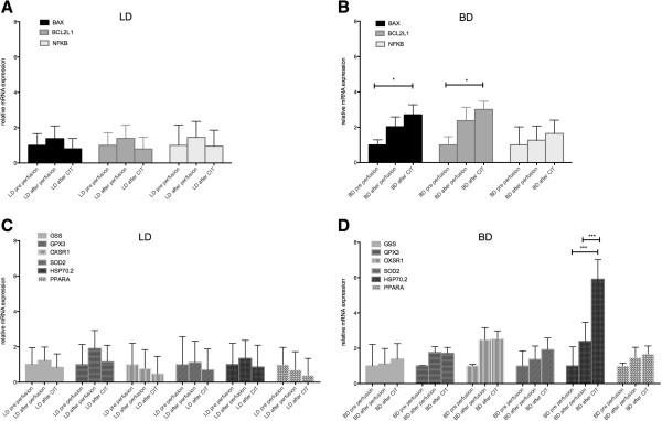
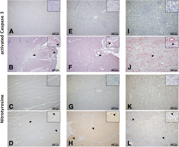
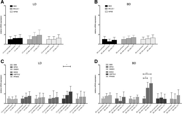
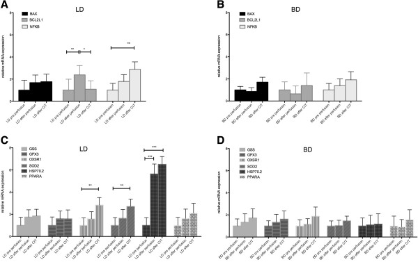
Results: In heart tissue of BD BAX, BCL2L1 and HSP 70.2 increased significantly after CIT. Only SOD2 was over-expressed after CIT in BD liver tissue. In kidney tissue, BCL2L1, NFKB, OXSR1, SOD2 and HSP 70.2 expression was significantly elevated in LD. Immunohistochemistry showed a significant increase in activated Caspase 3 and nitrotyrosine positive cells after CIT in BD in liver and in kidney tissue but not in heart tissue.
Conclusion: The up-regulation of protective and apoptotic genes seems to be divergent in the different organs in the BD and LD setting; however, immunohistochemistry revealed more apoptotic and nitrotyrosine positive cells in the BD setting in liver and kidney tissue whereas in heart tissue both BD and LD showed an increase.
Figures
References
-
- Terasaki PI, Cecka JM, Gjertson DW, Takemoto S. High survival rates of kidney transplants from spousal and living unrelated donors. N Engl J Med. 1995;333:333–336. - PubMed
-
- Herijgers P, Leunens V, Tjandra-Maga TB, Mubagwa K, Flameng W. Changes in organ perfusion after brain death in the rat and its relation to circulating catecholamines. Transplantation. 1996;62:330–335. - PubMed
-
- Dutkiewicz G, Domanski L, Binczak-Kuleta A, Pawlik A, Safranow K, Dziedziejko V, Wisniewska M, Ciechanowicz A, Ciechanowski K. Lack of association of polymorphisms 239 + 34A/C in the SOD1 gene and 47C/T in the SOD2 gene with delayed graft function and acute and chronic rejection of kidney allografts. Transplant Proc. 2009;41:3701–3703. - PubMed
-
- Pratschke S, Bilzer M, Grutzner U, Angele M, Tufman A, Jauch KW, Schauer RJ. Tacrolimus preconditioning of rat liver allografts impacts glutathione homeostasis and early reperfusion injury. J Surg Res. 2012;176:309–316. - PubMed
MeSH terms
Substances
LinkOut - more resources
Full Text Sources
Other Literature Sources
Research Materials
Miscellaneous
