

Hyperperfusion in progressive multifocal leukoencephalopathy is associated with disease progression and absence of immune reconstitution inflammatory syndrome
- PMID: 24088807
- PMCID: PMC3808691
- DOI: 10.1093/brain/awt268
Hyperperfusion in progressive multifocal leukoencephalopathy is associated with disease progression and absence of immune reconstitution inflammatory syndrome
Abstract
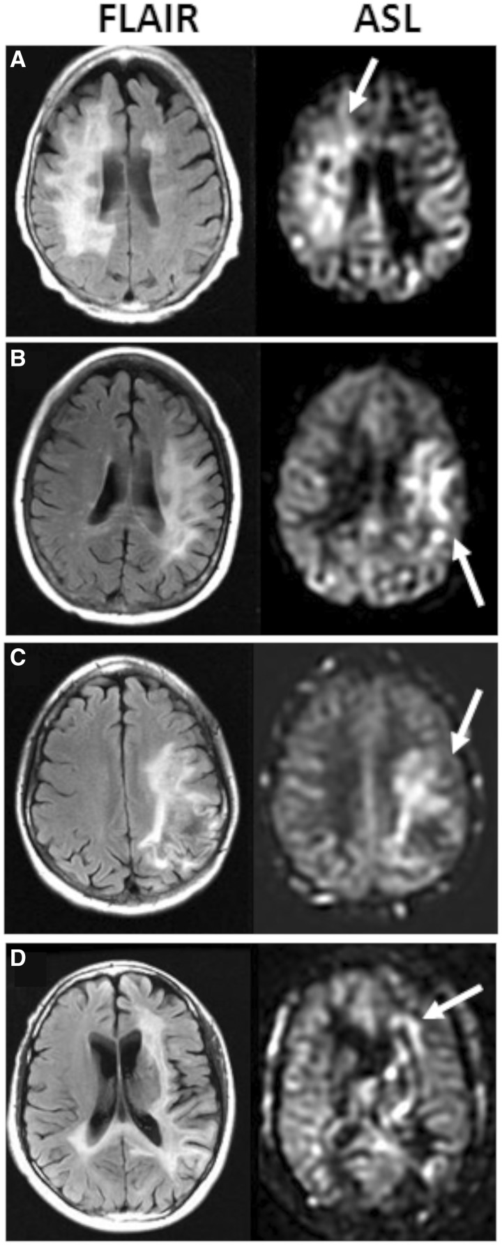
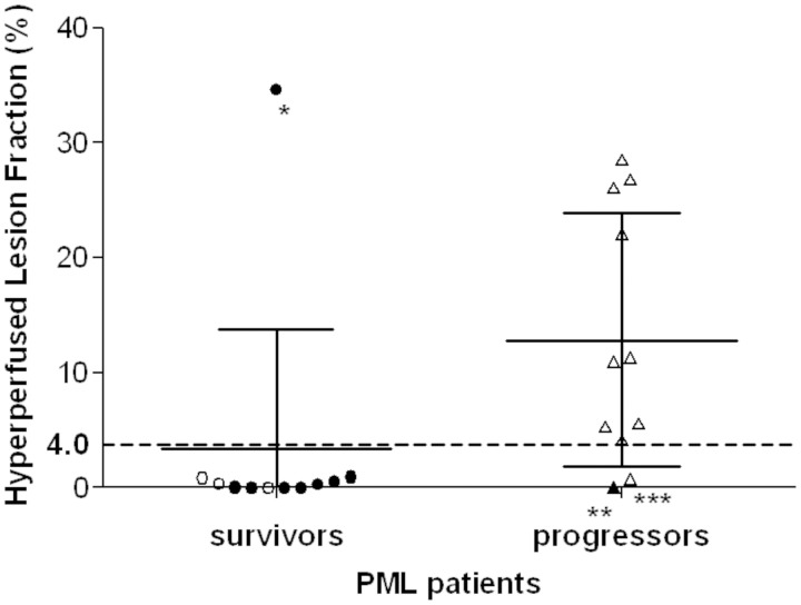
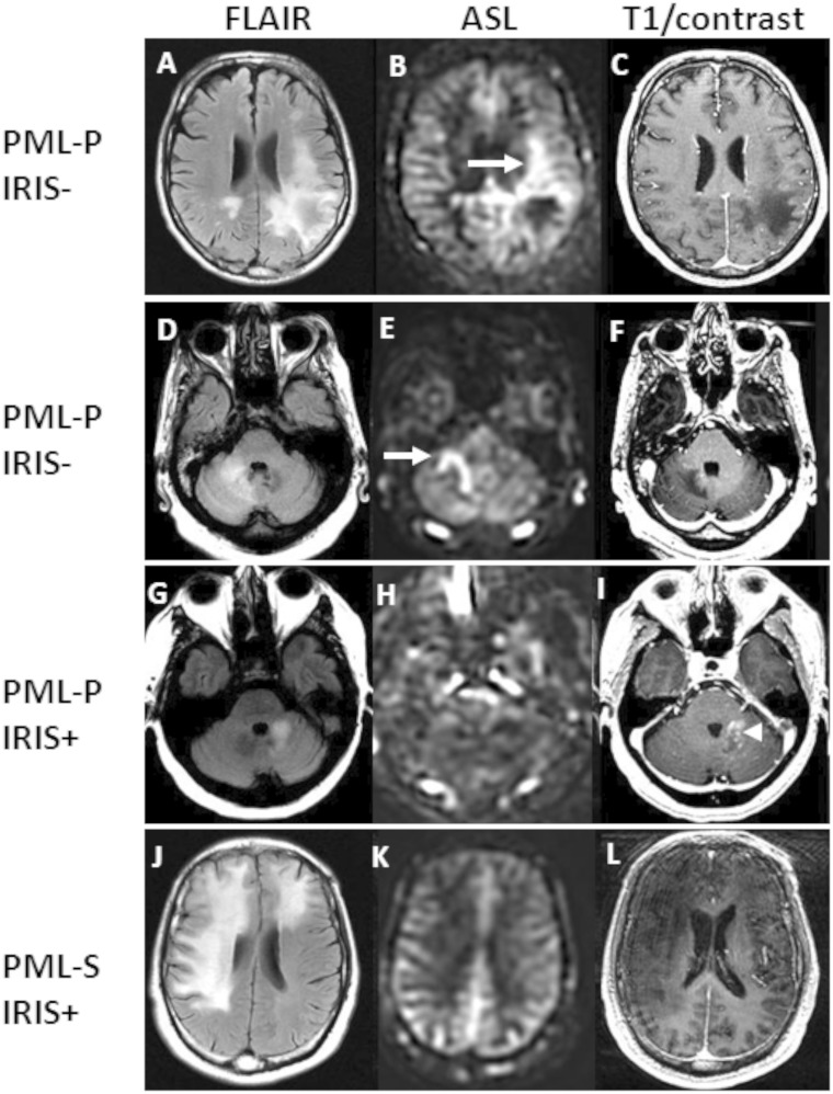
We sought to characterize perfusion patterns of progressive multifocal leukoencephalopathy lesions by arterial spin labelling perfusion magnetic resonance imaging and to analyse their association with immune reconstitution inflammatory syndrome, and survival. A total of 22 patients with progressive multifocal leukoencephalopathy underwent a clinical evaluation and magnetic resonance imaging of the brain within 190 days of symptom onset. The presence of immune reconstitution inflammatory syndrome was determined based on clinical and laboratory criteria. Perfusion within progressive multifocal leukoencephalopathy lesions was determined by arterial spin labelling magnetic resonance imaging. We observed intense hyperperfusion within and at the edge of progressive multifocal leukoencephalopathy lesions in a subset of subjects. This hyperperfusion was quantified by measuring the fraction of lesion volume showing perfusion in excess of twice normal appearing grey matter. Hyperperfused lesion fraction was significantly greater in progressive multifocal leukoencephalopathy progressors than in survivors (12.8% versus 3.4% P = 0.02) corresponding to a relative risk of progression for individuals with a hyperperfused lesion fraction ≥ 4.0% of 9.1 (95% confidence interval of 1.4-59.5). The presence of hyperperfusion was inversely related to the occurrence of immune reconstitution inflammatory syndrome at the time of scan (P = 0.03). Indeed, within 3 months after symptom onset, hyperperfusion had a positive predictive value of 88% for absence of immune reconstitution inflammatory syndrome. Arterial spin labelling magnetic resonance imaging recognized regions of elevated perfusion within lesions of progressive multifocal leukoencephalopathy. These regions might represent virologically active areas operating in the absence of an effective adaptive immune response and correspond with a worse prognosis.
Keywords: magnetic resonance imaging; neuroimmunology; neuroinflammation; perfusion imaging; progressive multifocal leukoencephalopathy.
Figures
Similar articles
-
Clinical and Radiological Characterization of Progressive Multifocal Leukoencephalopathy in HIV-Infected Patients: A Retrospective Analysis and Review of the Literature.Acta Med Port. 2015 May-Jun;28(3):286-96. doi: 10.20344/amp.5950. Epub 2015 Jun 30. Acta Med Port. 2015. PMID: 26421780 Review.
-
[Progressive multifocal leukoencephalopathy and natalizumab-related immune reconstitution inflammatory syndrome: the advantage of MRI].Rev Neurol (Paris). 2014 Jan;170(1):48-50. doi: 10.1016/j.neurol.2013.03.009. Epub 2013 Oct 17. Rev Neurol (Paris). 2014. PMID: 24139244 French. No abstract available.
-
The chameleon of neuroinflammation: magnetic resonance imaging characteristics of natalizumab-associated progressive multifocal leukoencephalopathy.Mult Scler. 2013 Dec;19(14):1826-40. doi: 10.1177/1352458513510224. Epub 2013 Nov 5. Mult Scler. 2013. PMID: 24192217 Review.
-
Severe Progressive Multifocal Leukoencephalopathy (PML) and Spontaneous Immune Reconstitution Inflammatory Syndrome (IRIS) in an Immunocompetent Patient.Front Immunol. 2019 May 28;10:1188. doi: 10.3389/fimmu.2019.01188. eCollection 2019. Front Immunol. 2019. PMID: 31191548 Free PMC article.
-
The spectrum of progressive multifocal leukoencephalopathy: a practical approach.Eur J Neurol. 2019 Apr;26(4):566-e41. doi: 10.1111/ene.13906. Epub 2019 Feb 8. Eur J Neurol. 2019. PMID: 30629326 Review.
Cited by
-
The Imaging of Localization Related Symptomatic Epilepsies: The Value of Arterial Spin Labelling Based Magnetic Resonance Perfusion.Korean J Radiol. 2018 Sep-Oct;19(5):965-977. doi: 10.3348/kjr.2018.19.5.965. Epub 2018 Aug 6. Korean J Radiol. 2018. PMID: 30174487 Free PMC article.
-
Tumefactive demyelination in a patient with relapsing-remitting MS on ocrelizumab.Neurol Neuroimmunol Neuroinflamm. 2019 Jun 26;6(5):e589. doi: 10.1212/NXI.0000000000000589. Print 2019 Sep. Neurol Neuroimmunol Neuroinflamm. 2019. PMID: 31454764 Free PMC article. No abstract available.
-
Arterial Spin Labeling technique and clinical applications of the intracranial compartment in stroke and stroke mimics - A case-based review.Neuroradiol J. 2022 Aug;35(4):437-453. doi: 10.1177/19714009221098806. Epub 2022 May 30. Neuroradiol J. 2022. PMID: 35635512 Free PMC article. Review.
-
Arterial Spin Labeling: Key Concepts and Progress Towards Use as a Clinical Tool.Magn Reson Med Sci. 2024 Jul 1;23(3):352-366. doi: 10.2463/mrms.rev.2024-0013. Epub 2024 Jun 14. Magn Reson Med Sci. 2024. PMID: 38880616 Free PMC article. Review.
-
Overview of MRI findings in progressive multifocal leukoencephalopathy.Jpn J Radiol. 2025 Jul 21. doi: 10.1007/s11604-025-01837-y. Online ahead of print. Jpn J Radiol. 2025. PMID: 40690111 Review.
References
-
- Abrams J. Nitrate delivery systems in perspective. A decade of progress. Am J Med. 1984;76:38–46. - PubMed
-
- Aiello S, Noris M, Piccinini G, Tomasoni S, Casiraghi F, Bonazzola S, et al. Thymic dendritic cells express inducible nitric oxide synthase and generate nitric oxide in response to self- and alloantigens. J Immunol. 2000;164:4649–58. - PubMed
-
- Alsop DC, Detre JA. Reduced transit-time sensitivity in noninvasive magnetic resonance imaging of human cerebral blood flow. J Cereb Blood Flow Metab. 1996;16:1236–49. - PubMed
-
- Alsop DC, Detre JA, Grossman M. Assessment of cerebral blood flow in Alzheimer's disease by spin-labeled magnetic resonance imaging. Ann Neurol. 2000;47:93–100. - PubMed
Publication types
MeSH terms
Grants and funding
LinkOut - more resources
Full Text Sources
Other Literature Sources
