. 2013 Nov 14;89(5):113.
doi: 10.1095/biolreprod.113.113159. Print 2013 Nov.

Retinoblastoma protein (RB1) controls fate determination in stem cells and progenitors of the mouse male germline

Affiliations

Retinoblastoma protein (RB1) controls fate determination in stem cells and progenitors of the mouse male germline

Qi-En Yang et al. Biol Reprod. .

Abstract

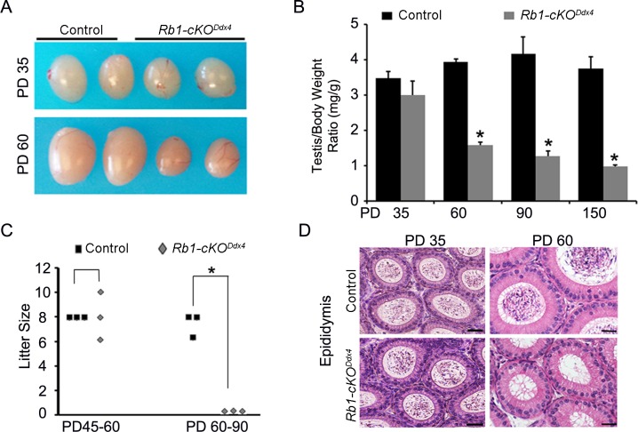
Continual spermatogenesis is the cornerstone of male fertility and relies on the actions of an undifferentiated spermatogonial population comprised of stem cells and progenitors. A foundational spermatogonial stem cell (SSC) pool is established during postnatal development that serves as a self-renewing reservoir from which progenitor spermatogonia arise that transiently amplify in number before committing to terminal differentiation. At present, the underlying molecular mechanisms governing these actions are undefined. Using conditional mutant mouse models, we investigated whether function of the undifferentiated spermatogonial population during postnatal life is influenced by the tumor suppressor protein RB1. Spermatogenesis initiates in mice with conditional inactivation of Rb1 in prospermatogonial precursors, but the germline is progressively lost upon aging due to impaired renewal of the undifferentiated spermatogonial population. In contrast, continual spermatogenesis is sustained following Rb1 inactivation in progenitor spermatogonia, but some cells transform into a carcinoma in situ-like state. Furthermore, knockdown of Rb1 abundance within primary cultures of wild-type undifferentiated spermatogonia impairs maintenance of the SSC pool, and some cells are invasive of the basement membrane after transplant into recipient testes, indicating acquisition of tumorigenic properties. Collectively, these findings indicate that RB1 plays an essential role in establishment of a self-renewing SSC pool and commitment to the spermatogenic lineage within progenitor spermatogonia.

Keywords: spermatogenesis; spermatogonia; spermatogonial stem cells; stem cells; testis.


Figures

FIG. 1
FIG. 1
Impact of Rb1 inactivation in prospermatogonia on male fertility in adulthood. A) Testes from control and Rb1-cKODdx4 mice at PD 35 and 60. B) Testis/body weight ratio (mg/g) of control and Rb1-cKODdx4 testes at PD 35, 60, 90, and 150. C) Fertility assessment of Rb1-cKODdx4 and control male mice based on number of pups sired after mating with wild-type females at PD 45–60 and 60–90. D) Cross-sections from caput epididymis of control and Rb1-cKODdx4 mice at PD 35 and 60. At PD 60, epididymal tubules from Rb1-cKODdx4 mice were devoid of spermatozoa. All the quantitative data are presented as mean ± SEM for three different mice of each genotype. Bars = 20 μm, and *denotes significantly different at P < 0.05.
FIG. 2
FIG. 2
Impact of Rb1 inactivation in prospermatogonia on spermatogenesis in adulthood. A) Hematoxylin and eosin-stained cross-sections from testes of control and Rb1-cKODdx4 mice at PD 35, 60, 90, and 150. Seminiferous tubules with disrupted spermatogenesis are noticeable at PD 60, including those with apparent Sertoli-cell-only (SCO) phenotype (asterisks). B) Seminiferous tubule cross-sections with apparent SCO phenotype from testes of Rb1-cKODdx4 mice and normal spermatogenesis from testes of control mice at PD 60 stained with hematoxylin and eosin or by immunofluorescence for the Sertoli cell marker SOX9 and germ cell marker GCNA1. DNA is stained with DAPI. Lack of staining for GCNA1 but staining for SOX9 confirms an SCO phenotype. C, D) Quantification of seminiferous tubule cross-sections from testes of Rb1-cKODdx4 mice containing degenerated spermatogenesis (C) and an SCO phenotype (D) at PD 35, 60, 90, and 150. Data are the mean ± SEM percentage of all the seminiferous tubule cross-sections for three different mice. Bars = 50 μm (A) and 20 μm (B).
FIG. 3
FIG. 3
Impaired renewal of the undifferentiated spermatogonial population during neonatal development in the absence of RB1. A) Immunofluorescent staining for the germ cell marker GCNA1 and Sertoli cell marker SOX9 in cross-sections of testes from control and Rb1-cKODdx4 mice at PD 0 and 6. B) Quantification of germ cells (GCNA1+) per seminiferous tubule cross-section from testes of Rb1-cKODdx4 and control mice at PD 0 and 6. C) Immunofluorescent staining for GCNA1 and EdU in cross-sections of testes from control and Rb1-cKODdx4 mice at PD 1 and 2. D) Immunofluorescent staining for the undifferentiated spermatogonial markers LIN28 and ZBTB16 in cross-sections of testes from control and Rb1-cKODdx4 mice at PD 6 and 10. E) Quantification of ZBTB16+/LIN28+ germ cells per seminiferous tubule cross-section from testes of control and Rb1-cKODdx4 mice at PD 6 and 10. F) Immunofluorescent staining for ZBTB16+/EdU+ and ZBTB16+/TUNEL+ cells in cross-sections of seminiferous tubules from testes of control and Rb1-cKODdx4 mice at PD 6. G, H) Quantification of ZBTB16+/EdU+ spermatogonia (G) and ZBTB16+/TUNEL+ spermatogonia (H) per seminiferous tubules cross-section of testes from control and Rb1-cKODdx4 mice at PD 6. I) Immunofluorescent staining for expression of the undifferentiated spermatogonial marker ZBTB16 and differentiating spermatogonial marker KIT in cross-sections of seminiferous tubules from testes of control and Rb1-cKODdx4 mice at PD 10. For all the images, DAPI was used to stain DNA, and bars = 20 μm. All the quantitative data are presented as mean ± SEM for three different mice of each genotype, and *denotes significantly different at P < 0.05.
FIG. 4
FIG. 4
Impaired spermatogenic lineage commitment of progenitor spermatogonia in the absence of RB1. A) Testes from control and Rb1-cKONeurog3 mice. A cyst (arrow) is evident in the testis from the Rb1-cKONeurog3 mouse. B) Cross-sections from testes of adult control and Rb1-cKONeurog3 mice stained with hematoxylin and eosin. Seminiferous tubules containing disrupted spermatogenesis (arrow) are evident in addition to the dilated rete testis region (asterisk). C) Hematoxylin-stained cross-section from the rete testis region of an Rb1-cKONeurog3 mouse. D) Hematoxylin and eosin-stained cross-sections from caput epididymis associated with normal and cyst containing testes of an Rb1-cKONeurog3 mouse at PD 60. Spermatozoa are evident within epididymal tubules associated with the normal testis but absent within tubules associated with the cyst-containing testis. E) Hematoxylin and eosin-stained cross-sections of seminiferous tubules from testes of control mice and cyst containing testes from Rb1-cKONeurog3 and Rb1-cKOStra8 mice. Normal spermatogenesis is evident within seminiferous tubules from control mice, and tubules from Rb1-cKONeurog3 and Rb1-cKOStra8 mice contain disrupted spermatogenesis and abnormal germ cells with a CIS-like morphology (arrow). F) Immunofluorescent staining for markers of Sertoli cells (SOX9), germ cells (GCNA1), undifferentiated spermatogonia (ZBTB16/LIN28), premeiotic spermatogonia (STRA8), and meiotic spermatocytes (gH2A.X) in cross- sections of seminiferous tubules from normal and cyst containing testes of an Rb1-cKONeurog3 mouse. G) Alkaline phosphatase (AP) staining of cross- sections from normal and cyst testes of an Rb1-cKONeurog3 mouse. AP+ cells are evident within seminiferous tubules of the cyst containing testis (arrow). H) Immunofluorescent staining for GCNA1+ and TUNEL+ cells in cross-sections of seminiferous tubules from normal and cyst testes of an Rb1-cKONeurog3 mouse. Bars = 2 mm (A), 50 μm (B), or 20 μm (CH).
FIG. 5
FIG. 5
Impaired maintenance of the SSC pool and spermatogenic lineage commitment within primary cultures of wild-type undifferentiated spermatogonia following transient reduction of RB1. A) Recipient mouse testes 2 mo after transplantation with cultured undifferentiated spermatogonia established from Rosa-LacZ-expressing donor mice 6 days after treatment with control or Rb1 siRNA. B) Quantitative comparison of SSC numbers in primary cultures of undifferentiated spermatogonia 6 days after treatment with control or Rb1 siRNA based on donor-derived colonies of spermatogenesis and normalization to 105 cells injected. Data are mean ± SEM for three different cultures and 12 total recipient testes for each treatment. Asterisk (*) denotes significantly different at P < 0.05. C) Total germ cell number in primary cultures of undifferentiated spermatogonia 6 days after treatment with control or Rb1 siRNA. Data are mean ± SEM for three different cultures. D) Recipient mouse seminiferous tubules 60 days after transplantation of cultured spermatogonia treated with control or Rb1 siRNA for 6 days. Arrow indicates abnormal colony formation from Rb1 siRNA-treated cells. E) Cross-sections of recipient mouse testes stained for LacZ-expressing donor cells 60 days after transplantation of control or Rb1 siRNA-treated germ cells into seminiferous tubules. Complete colonies of spermatogenesis were established from both control and Rb1 siRNA-treated SSCs (asterisks); however, some Rb1 siRNA-treated germ cells were invasive of the basement membrane and colonized the interstitial tissue (arrows). F) Quantitative comparison of cells in S-phase within primary cultures of undifferentiated spermatogonia 48 h posttreatment with control or Rb1 siRNA based on EdU incorporation. Data are mean ± SEM for three different cultures, and *denotes significantly different at P < 0.05. G) Quantitative real-time RT-PCR analysis for Ccnd1, Ccnd2, Ccnd3, Ccne1, and Ccne2 transcript abundance in primary cultures of undifferentiated spermatogonia 48 h after treatment with control or Rb1 siRNA. Data are mean ± SEM fold-difference of Rb1 siRNA to control, and *denotes significantly different at P < 0.05. Bars = 2 mm (A) or 20 μm (DE).

Similar articles

Cited by

References

    1. Oatley JM, Brinster RL. Regulation of spermatogonial stem cell self-renewal in mammals Annu Rev Cell Dev Biol 2008. 24 263– 286 - PMC - PubMed
    1. De Rooij DG. Regulation of the proliferation of spermatogonial stem cells J Cell Sci Suppl 1988. 10 181– 194 - PubMed
    1. Oatley JM, Brinster RL. The germline stem cell niche unit in mammalian testes Physiol Rev 2012. 92 577– 595 - PMC - PubMed
    1. Yoshinaga K, Nishikawa S, Ogawa M, Hayashi S, Kunisada T, Fujimoto T. Role of c-kit in mouse spermatogenesis: identification of spermatogonia as a specific site of c-kit expression and function Development 1991. 113 689– 699 - PubMed
    1. Yang QE, Racicot KE, Kaucher AV, Oatley MJ, Oatley JM. MicroRNAs 221 and 222 regulate the undifferentiated state in mammalian male germ cells Development 2013. 140 280– 290 - PMC - PubMed

Publication types

Substances