. 2014 Jan 1;306(1):R10-22.
doi: 10.1152/ajpregu.00240.2013. Epub 2013 Oct 2.

Paradoxical effects of heme arginate on survival of myocutaneous flaps

Affiliations

Paradoxical effects of heme arginate on survival of myocutaneous flaps

Marie-Claire Edmunds et al. Am J Physiol Regul Integr Comp Physiol. .

Abstract

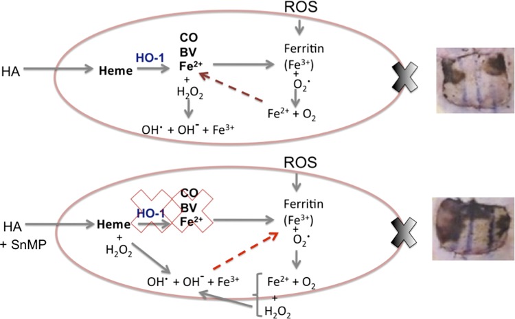
Ischemia reperfusion injury (IRI) contributes to partial flap and solid organ transplant failure. Heme-oxygenase 1 (HO-1) is an inducible, cytoprotective enzyme which protects against IRI in solid organ transplant models. Heme arginate (HA), a HO-1 inducer, is a promising, translatable, preconditioning agent. This study investigated the effects of preconditioning with HA on the clinical outcome of a myocutaneous IRI model. Forty male Lewis rats were randomized to intravenously receive 1) Control-NaCl, 2) HA, 3) HA and tin mesoporphyrin (SnMP), a HO-1 inhibitor; and 4) SnMP alone. Twenty-four hours later, an in situ transverse rectus abdominis myocutaneous flap was performed under isoflurane anesthesia. Viability of flaps was measured clinically and by laser-Doppler perfusion scanning. In vitro work on human epidermal keratinocytes (HEKa) assessed the effects of HA, SnMP, and the iron chelator desferrioxamine on 1) cytotoxicity, 2) intracellular reactive oxygen species (ROS) concentration, and 3) ROS-mediated DNA damage. In contrast to our hypothesis, HA preconditioning produced over 30% more flap necrosis at 48 h compared with controls (P = 0.02). HA-containing treatments produced significantly worse flap perfusion at all postoperative time points. In vitro work showed that HA is cytotoxic to keratinocytes. This cytotoxicity was independent of HO-1 and was mediated by the generation of ROS by free heme. In contrast to solid organ data, pharmacological preconditioning with HA significantly worsened clinical outcome, thus indicating that this is not a viable approach in free flap research.

Keywords: free tissue transfer; heme arginate; heme-oxygenase-1; ischemia reperfusion injury; myocutaneous flap.


Figures

Fig. 1.
Fig. 1.
Western blots from skin (A), muscle (B), fat (C), spleen (D), kidney (E), and liver (F). Protein lysates were obtained from tissues harvested 24 h after administration of 0, 5, 15, and 30 mg/kg of HA via intravenous injection. The upper bands are β-actin (42 kDa), a loading control, and the lower bands are HO-1 (32 kDa). Of all the tissues assessed, only the skin and spleen show baseline HO-1 activity. A dose-related increase in HO-1 was found in all the tissues assessed. G: HO-1 activity assay results (n = 3 per group). Animals received intravenous preconditioning with control = NaCl; HA (30 mg/kg); heme arginate (HA) + tin mesoporphyrin (SnMP) (30 mg/kg HA and 40 μmol/kg SnMP); and SnMP (40 μmol/kg) alone. After 24 h, the animals were culled, tissue was harvested, and samples were prepared from homogenized livers. HA administration resulted in a significant increase of HO-1 [F(3, 8) = 228, P < 0.0001; r2 = 0.99]. This activity was abrogated by coadministration of SnMP. Statistically significant differences between preconditioning groups are indicated as follows: ***P < 0.001, as established be one-way ANOVA and Tukey's post hoc test.
Fig. 2.
Fig. 2.
AD: HO-1-positive staining within the skin with ×100 magnification (×400, insets) for control = NaCl (A), HA (30 mg/kg) (B), HA + SnMP (30 mg/kg HA and 40 μmol/kg SnMP) (C), and SnMP (40 μmol/kg) alone (D). E: colocalization of HO-1 (red) and pan-macrophage marker CD68 (green) at ×400 confocal magnification. White arrows indicate cells in which CD68 and HO-1 were colocalized. Blue arrow indicates HO-1-positive cells with no concurrent CD68 staining. F–H: black and white images of the three separate channels, which make up composite image (E): DAPI (F), CD68 (G), and HO-1 (H).
Fig. 3.
Fig. 3.
Representative results of necrosis and perfusion 48 h postoperatively. Animals were randomized to receive either Control-NaCl, HA (30 mg/kg), HA + SnMP (30 mg/kg HA + 40 μmol/kg SnMP), and SnMP (40 μmol/kg) alone by intravenous injection. After 24 h, these animals underwent the transverse rectus abdominis myocutaneous (TRAM) procedure. The preconditioning groups were control, HA, HA + SnMP, and SnMP. The upper row are high-resolution images from which percentage area necrosis was calculated. The lower row shows the corresponding laser-Doppler imaging scans. Perfusion is given in perfusion units, and each unit is assigned a color. The scale is shown below the images.
Fig. 4.
Fig. 4.
A: mean percentage area flap necrosis at 48 h following TRAM flap procedure. A one-way ANOVA between the pretreatment groups on this outcome measure was significant, P < 0.0001. Tukey's post hoc comparison of the mean values of these groups showed a significant increase in flap necrosis in both the HA and HA + SnMP-pretreated groups compared with control and SnMP-pretreated animals as denoted by *P < 0.05, **P < 0.01, and ***P < 0.001; n = 10. Values are expressed as means ± SE. B–D: hematoxylin-and-eosin histological classification of injury composite score. Animals were administered the following: Control-NaCl, HA (30 mg/kg), HA + SnMP (30 mg/kg HA and 40 μmol/kg SnMP), and SnMP (40 μmol/kg) alone by intravenous injection. After 24 h, these animals underwent the TRAM procedure. Methyl Carnoy's solution-fixed, paraffin-embedded sections were harvested 48 h after the animals underwent sham surgery (n = 4) (B), ischemia reperfusion injury (IRI)-Zone IV (n = 10) (C), and IRI- Zone I (n = 10) (D). Results were analyzed by one-way ANOVA and revealed significant differences between the treatment groups on this composite score outcome: [F(4,13) = 4.98, P = 0.0117] (B), [F(4, 33) = 21.9, P < 0.0001] (C), and [F(4,33) = 29.4, P < 0.0001] (D). The results of Tukey's post hoc test are denoted by *P < 0.05, **P < 0.01, and ***P < 0.001. Values are expressed as means ± SE.
Fig. 5.
Fig. 5.
Mean flap perfusion as assessed by laser-Doppler imaging for the four preconditioning treatments: preoperatively (preop), immediately postoperatively (postop), at 24 h, and 48 h after the TRAM procedure. A significant difference between the pretreatment groups on this outcome measure was found by one-way ANOVA at all of the postoperative time points: postop (P = 0.0011), 24 h (P = 0.0002), and 48 h (P = 0.0004). Tukey's post hoc comparison test showed a significantly reduced perfusion in flaps treated with HA or HA + SnMP compared with controls at 24 and 48 h as indicated by *P < 0.05, **P < 0.01, and ***P < 0.001; n = 10. Values are expressed as means ± SE.
Fig. 6.
Fig. 6.
Mean cell viability as assessed by methylthiazolyldiphenyl-tetrazolium bromide (MTT) assay of human epidermal keratinocytes (HEKa) cells following 24 h of culture in the 12 preconditioning treatments. For clarity, only the results of control, 10 μmol HA, 10 μmol HA + 5 μmol SnMP, 5 μmol SnMP, 10 μmol HA + 500 μmol desferrioxamine (DF), 10 μmol HA + 50 μmol DF + 5 μmol SnMP, and 500 μmol DF are shown. A significant difference between the pretreatment groups on this outcome measure was found by one-way ANOVA, P < 0.0001. The seminal results of Tukey's test post hoc multiple comparisons are denoted by ns for not significant, **P < 0.01, and ***P < 0.001; n = 4. Values are expressed as means ± SE.
Fig. 7.
Fig. 7.
Mean cell viability as assessed by Vialight plus kit in HEKa cells following 24 h of culture in nine preconditioning treatments. A significant difference between the pretreatment groups on this outcome measure was found by one-way ANOVA, P < 0.0001. For simplicity, only the results of control, 10 μmol HA, 10 μmol HA + 5 μmol SnMP, 5 μmol SnMP, 10 μmol HA + 500 μmol DF, and 500 μmol DF are shown. The critical results from Tukey's post hoc comparison are denoted by ns for not significant, **P < 0.01, and ***P < 0.001; n = 3. Values are expressed as means ± SE. RLU, relative light units.
Fig. 8.
Fig. 8.
Geometric mean fluorescence, an output measure for intracellular reactive oxygen species (ROS) concentration, was measured by FACS following treatment with CMH2DCFDA. The 12 preconditioning treatments were found to produce significant effects on intracellular ROS concentration by one-way ANOVA, P < 0.0001. For comprehensibility, the results of control, 10 μmol HA, 10 μmol HA + 5 μmol SnMP, 5 μmol SnMP, 10 μmol HA + 500 μmol DF, and 500 μmol DF are shown. The results of Tukey's post hoc comparison are denoted by **P < 0.01, ***P < 0.001, ns, not significant; n = 3. Values are expressed as means ± SE.
Fig. 9.
Fig. 9.
8-Hydroxy-2′-deoxguanosine (8-OHdg), a measure for oxidative stress-specific DNA damage. The 12 preconditioning treatments were found to produce significant effects on 8-OHdg levels by one-way ANOVA, P < 0.0001. The results of Tukey's post hoc comparison are denoted by **P < 0.01 and ***P < 0.001; n = 3. Values are expressed as means ± SE.
Fig. 10.
Fig. 10.
Proposed mechanism of HA-mediated cell damage in the in vivo model. Top: upon administration of HA excess free heme results in the upregulation of HO-1 and production of CO, biliverdin (BV), and Fe2+. Both the free heme and Fe2+ can act as Fenton reactors to produce ROS. Fe2+ results in the expedient production of ferritin, which sequesters the reactive Fe2+ and oxidizes it to less reactive Fe3+. BV is rapidly converted by cytosolic biliverdin reductase to bilirubin, and both have powerful antioxidant activities to counteract the production of ROS from free heme and Fe2+. The scenario is complicated in the IRI model by the generation of ROS from reperfusion of previously ischemic tissues. This further strains the cells' intrinsic antioxidant capacity and may lead to the release of Fe3+ from ferritin, further exacerbating the production of ROS. This leads to necrosis (image, top, right). In the HA + SnMP-treated animals (bottom), the situation is worsened by the inhibition of HO-1. This further decreases the cells' ability to quench the ROS produced from the heme load from administration of HA and the IRI. This results in more extensive tissue damage (image, bottom, right). HA, heme arginate; HO-1, heme-oxygenase-1; CO, carbon monoxide; BV, biliverdin; Fe2+, ferrous iron; Fe3+, ferric iron; H2O2, hydrogen peroxide; OH, hydroxyl ion; OH·, hydroxyl radical; ROS, reactive oxygen species; O2, oxygen; SnMP, tin mesoporphyrin.

Similar articles

Cited by

References

    1. Andreas M, Schmid AI, Doberer D, Schewzow K, Weisshaar S, Heinze G, Bilban M, Moser E, Wolzt M. Heme arginate improves reperfusion patterns after ischemia: a randomized, placebo-controlled trial in healthy male subjects. J Cardiovasc Magn Reson 14: 55, 2012 - PMC - PubMed
    1. Arosio P, Ingrassia R, Cavadini P. Ferritins: a family of molecules for iron storage, antioxidation and more. Biochim Biophys Acta 1790: 589–599, 2009 - PubMed
    1. Attuwaybi BO, Kozar RA, Moore-Olufemi SD, Sato N, Hassoun HT, Weisbrodt NW, Moore FA. Heme oxygenase-1 induction by hemin protects against gut ischemia/reperfusion injury. J Surg Res 118: 53–57, 2004 - PubMed
    1. Bach FH. Heme oxygenase-1: a therapeutic amplification funnel. FASEB J 19: 1216–1219, 2005 - PubMed
    1. Balla J, Vercellotti GM, KN, Yachie A, Nagy E, Eaton JW, Balla G. Haem, haem oxygenase and ferritin in vascular endothelial cell injury. Nephrol Dial Transpl 18: v8–v12, 2003 - PubMed

Publication types

LinkOut - more resources