

α-Synuclein in cutaneous autonomic nerves
- PMID: 24089386
- PMCID: PMC3806913
- DOI: 10.1212/WNL.0b013e3182a9f449
α-Synuclein in cutaneous autonomic nerves
Abstract
Objective: To develop a cutaneous biomarker for Parkinson disease (PD).
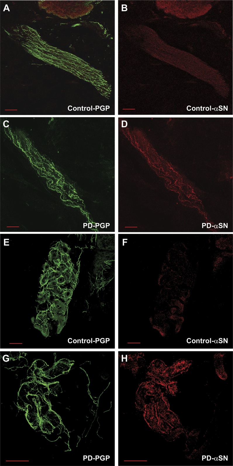
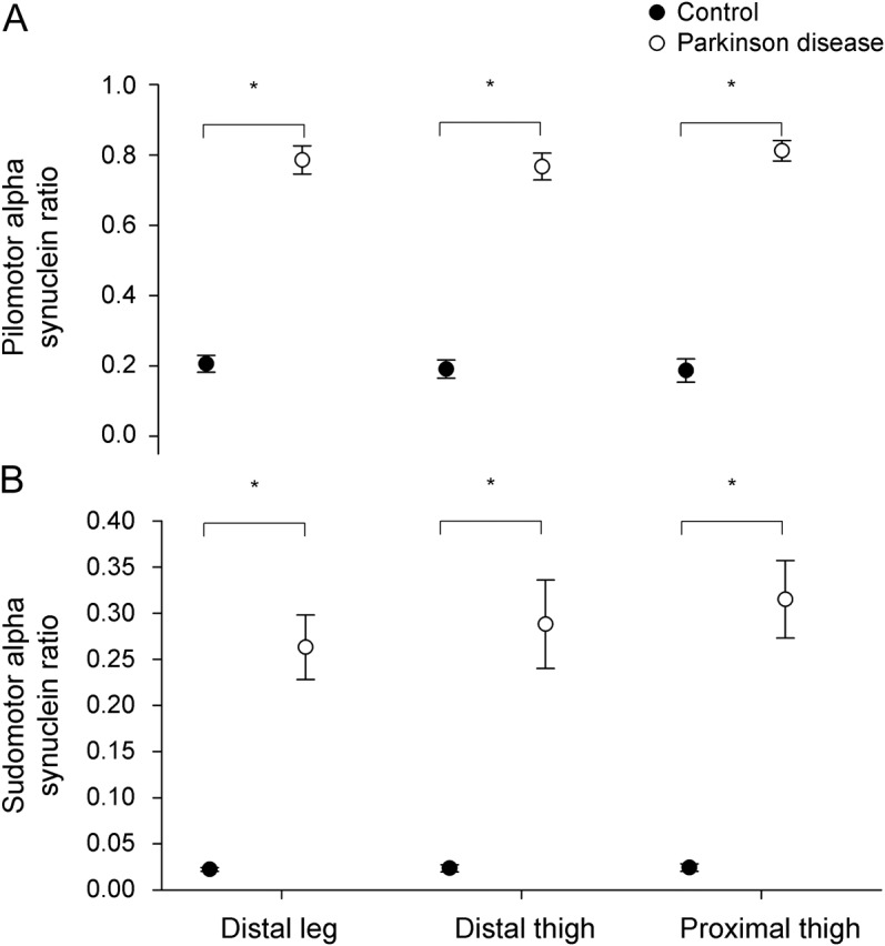
Methods: Twenty patients with PD and 14 age- and sex-matched control subjects underwent examinations, autonomic testing, and skin biopsies at the distal leg, distal thigh, and proximal thigh. α-Synuclein deposition and the density of intraepidermal, sudomotor, and pilomotor nerve fibers were measured. α-Synuclein deposition was normalized to nerve fiber density (the α-synuclein ratio). Results were compared with examination scores and autonomic function testing.
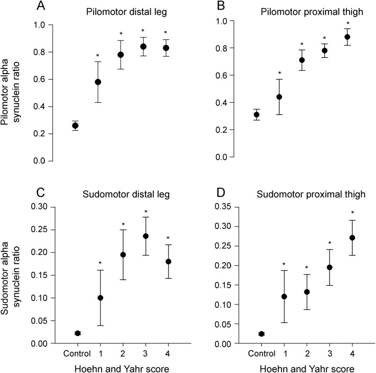
Results: Patients with PD had a distal sensory and autonomic neuropathy characterized by loss of intraepidermal and pilomotor fibers (p < 0.05 vs controls, all sites) and morphologic changes to sudomotor nerve fibers. Patients with PD had greater α-synuclein deposition and higher α-synuclein ratios compared with controls within pilomotor nerves and sudomotor nerves (p < 0.01, all sites) but not sensory nerves. Higher α-synuclein ratios correlated with Hoehn and Yahr scores (r = 0.58-0.71, p < 0.01), with sympathetic adrenergic function (r = -0.40 to -0.66, p < 0.01), and with parasympathetic function (r = -0.66 to -0.77, p > 0.01).
Conclusions: We conclude that α-synuclein deposition is increased in cutaneous sympathetic adrenergic and sympathetic cholinergic fibers but not sensory fibers of patients with PD. Higher α-synuclein deposition is associated with greater autonomic dysfunction and more advanced PD. These data suggest that measures of α-synuclein deposition in cutaneous autonomic nerves may be a useful biomarker in patients with PD.
Figures
Comment in
-
Is α-synuclein rising to the surface as a diagnostic biomarker for Parkinson disease?Neurology. 2013 Oct 29;81(18):1568-9. doi: 10.1212/WNL.0b013e3182a9f5ab. Epub 2013 Oct 2. Neurology. 2013. PMID: 24089391
References
-
- Dickson DW, Braak H, Duda JE, et al. Neuropathological assessment of Parkinson's disease: refining the diagnostic criteria. Lancet Neurol 2009;8:1150–1157 - PubMed
-
- Navarro-Otano J, Gelpi E, Mestres CA, et al. Alpha-synuclein aggregates in epicardial fat tissue in living subjects without parkinsonism. Parkinsonism Relat Disord 2013;19:27–31 - PubMed
-
- Michell AW, Luheshi LM, Barker RA. Skin and platelet alpha-synuclein as peripheral biomarkers of Parkinson's disease. Neurosci Lett 2005;381:294–298 - PubMed
-
- Ikemura M, Saito Y, Sengoku R, et al. Lewy body pathology involves cutaneous nerves. J Neuropathol Exp Neurol 2008;67:945–953 - PubMed
Publication types
MeSH terms
Substances
Grants and funding
LinkOut - more resources
Full Text Sources
Other Literature Sources
Medical