. 2013 Dec;143(12):1882-8.
doi: 10.3945/jn.113.184457. Epub 2013 Oct 2.

Gastric and colonic zinc transporter ZIP11 (Slc39a11) in mice responds to dietary zinc and exhibits nuclear localization

Affiliations

Gastric and colonic zinc transporter ZIP11 (Slc39a11) in mice responds to dietary zinc and exhibits nuclear localization

Alyssa B Martin et al. J Nutr. 2013 Dec.

Abstract

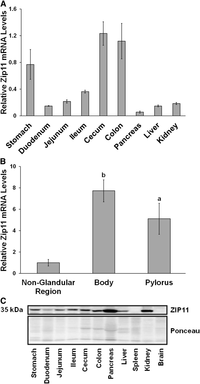
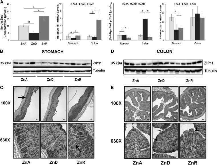
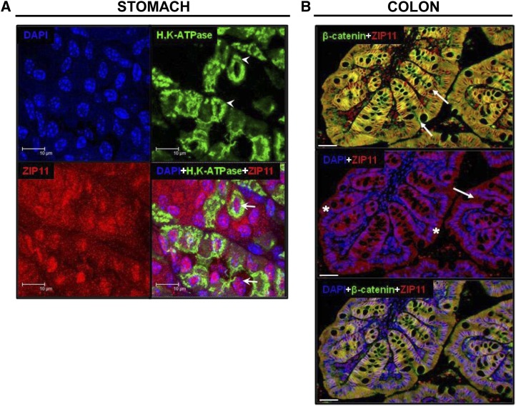
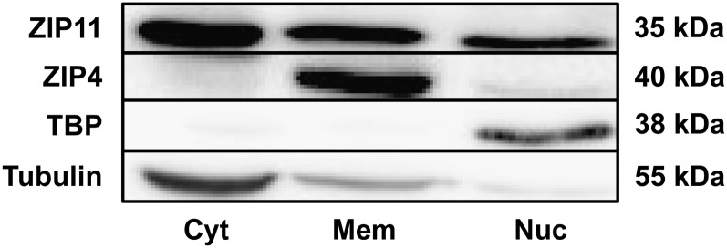
Zinc transporters have been characterized to further understand the absorption and metabolism of dietary zinc. Our goal was to characterize zinc transporter Slc39a11 (ZIP11) expression and its subcellular localization within cells of the murine gastrointestinal tract of mice and to determine if dietary zinc regulates ZIP11. The greatest ZIP11 expression was in the stomach, cecum, and colon. Both Zip11 mRNA and ZIP11 protein were shown to be downregulated during dietary zinc restriction (<1 mg Zn/kg) in the murine stomach tissue but were unaffected in the colon. Acute repletion with zinc did not restore Zip11 mRNA levels in the stomach. Immunohistochemistry (IHC) revealed high ZIP11 levels in the lower regions of gastric glands and parietal cells of the stomach. IHC analysis of the colon showed a marked ZIP11 abundance within the cytoplasm of the colonic epithelial cells. IHC also showed an increase in ZIP11 expression in the colon during zinc restriction. There is a robust abundance of ZIP11 in the nuclei of cells of both stomach and colon. Our experiments suggest that when dietary zinc intake is compromised, the colon may increase zinc transporter expression to improve the efficiency for absorption via increased expression of specific zinc transporters, including ZIP11 and also zinc transporter Slc39a4. In conclusion, ZIP11 is highly expressed within the murine stomach and colon and appears to be partially regulated by dietary zinc intake within these tissues. ZIP11 may play a specialized role in zinc homeostasis within these tissues, helping to maintain mucosal integrity and function.


Conflict of interest statement

Author disclosures: A. B. Martin, T. B. Aydemir, G. J. Guthrie, D. A. Samuelson, S.-M. Chang, and R. J. Cousins, no conflicts of interest.

Figures

FIGURE 1
FIGURE 1
Predicted topology for murine ZIP11 showing 6 transmembrane-spanning regions. The light gray box indicates the variable region of the isoforms. The dark gray box indicates the region used to produce our ZIP11 antibody. ZIP11, zinc transporter SLC39A11.
FIGURE 2
FIGURE 2
Tissue distribution of the Zip11 gene transcripts and ZIP11 protein in C57BL/6 mice. RNA was extracted from tissues and used for qPCR analysis of Zip11 mRNA. (A) Zip11 mRNA tissue distribution with pancreatic expression set to 1. (B) Zip11 mRNA expression distribution in the 3 regions of the mouse stomach. The nonglandular region expression was set to 1. Values were normalized to 18S. Values are means ± SDs, n = 4–5 for each tissue sample. Significant differences compared to the nonglandular region are indicated (a, P < 0.05; b, P < 0.01). (C) Western blots of total tissue lysates. ZIP11 is shown as a 35-kDa band. Ponceau staining from the blot was used as the loading control. ZIP11, zinc transporter SLC39A11.
FIGURE 3
FIGURE 3
Dietary ZnD and ZnR produce changes in ZIP11 expression in the stomach and colon compared with mice fed a ZnA diet. (A) Serum zinc concentration and relative expression of MT mRNA, Zip4 mRNA, and Zip11 mRNA in stomach and colon. mRNA levels for ZnA were set to 1. Data are expressed as means ± SDs, n = 3–5. Significant differences are indicated (a, P < 0.05; b, P < 0.01; c, P < 0.001; d, P < 0.0001). (B) Representative Western blot of stomach tissue lysate showing an abundance of the ZIP11 protein at 35 kDa, normalized to tubulin. (C) Immunoperoxidase staining of ZIP11 in the mouse stomach, visualized with a light microscope using 100× and 630× magnification. The stomach shows strong ZIP11 staining (dark gray) in the ZnA samples with a decrease in the ZnD samples. Black arrows indicate strong ZIP11 staining in the chief cell region and parietal cells of the gastric glands. ZIP11 staining returned to comparable levels seen in ZnA samples in the stomach after ZnR. (D) Representative Western blot of colon lysate showing abundance of the ZIP11 protein with tubulin as the loading control. (E) Immunoperoxidase staining of ZIP11 in the mouse colon is visualized at 100× and 630× magnification. The colon samples show an increase in staining in the ZnD samples compared with ZnA samples, and the staining remained the same after 24 h repletion. Black arrowheads indicate colonic crypts. Black lines in the 630× images represent 25 μm and those in the 100× images represent 50 μm. MT, Metallothionein; ZIP4, zinc transporter Slc39a4; ZIP11, zinc transporter SLC39A11; ZnA, zinc adequate; ZnD, zinc depletion; ZnR, acute zinc repletion.
FIGURE 4
FIGURE 4
IF imaging of ZIP11 in the mouse stomach and colon. (A) IF images of stomach are shown at 63× with 4× zoom. The tiled figure shows individual and merged fluorescence images. Images show the nuclei marker, DAPI (blue), the parietal cell marker, H,K-ATPase (green) (white arrowheads), and ZIP11, indicated by the red fluorescence. A merged image of all 3 antibodies indicated a localization of ZIP11 to the cytoplasm (red) and nuclei (pink) (white arrows) of gastric cells. The white bars represent 10 μm. (B) IF images of ZIP11 in the mouse colon are shown at 20×. The colonic folds are presented at high magnification to show the invaginations (colonic crypts) indicated by white arrows. The top merged image of ZIP11 (red) and β-catenin (green) indicates colocalization (yellow) of ZIP11 at the colonic epithelial cell plasma membrane. The middle merged image of ZIP11 (red) and DAPI (blue) shows colocalization (purple) of ZIP11 to the colonic epithelial cell nuclei (white asterisks). The bottom panel shows a merged image of ZIP11, β-catenin, and DAPI. This image indicates the localization of ZIP11 both at the colonic epithelial plasma membrane (yellow) and in the nuclei (purple). The white bars represent 25 μm. IF images were obtained using a laser spinning disc confocal microscope. DAPI, 4′,6-diamidino-2-phenylindole; IF, immunofluorescence; ZIP11, zinc transporter SLC39A11.
FIGURE 5
FIGURE 5
Western-blot analysis of ZIP11 from the subcellular fractionation of mouse colonic mucosal scrapings. Mucosal lysate was separated into a cytoplasmic fraction (Cyt), membrane fraction (Mem), and nuclear fraction (Nuc). ZIP11 was present in all three fractions. Controls for each fraction are shown: ZIP4 (mem), TBP (nuc), and tubulin (cyt). TBP, TATA binding protein; Zip4, zinc transporter Slc39a4; ZIP11, zinc transporter SLC39A11.

References

    1. Lichten LA, Cousins R. Mammalian zinc transporters: nutritional and physiologic regulation. Annu Rev Nutr. 2009;29:153–76. - PubMed
    1. Wang K, Zhou B, Kuo Y, Zemansky J, Gitschier J. A novel member of a zinc transporter family is defective in acrodermatitis enteropathica. Am J Hum Genet. 2002;71:66–73. - PMC - PubMed
    1. Wang F, Kim BE, Petris MJ, Eide DJ. The mammalian Zip5 protein is a zinc transporter that localizes to the basolateral surface of polarized cells. J Biol Chem. 2004;279:51433–41. - PubMed
    1. Liuzzi JP, Bobo JA, Lichten LA, Samuelson DA, Cousins RJ. Responsive transporter genes within the murine intestinal-pancreatic axis form a basis of zinc homeostasis. Proc Natl Acad Sci USA. 2004;101:14355–60. - PMC - PubMed
    1. McMahon RJ, Cousins RJ. Regulation of the zinc transporter ZnT-1 by dietary zinc. Proc Natl Acad Sci USA. 1998;95:4841–6. - PMC - PubMed

Publication types