. 2013 Dec;87(24):13354-69.
doi: 10.1128/JVI.02465-13. Epub 2013 Oct 2.

Evasion of superinfection exclusion and elimination of primary viral RNA by an adapted strain of hepatitis C virus

Affiliations

Evasion of superinfection exclusion and elimination of primary viral RNA by an adapted strain of hepatitis C virus

Brian Webster et al. J Virol. 2013 Dec.

Abstract

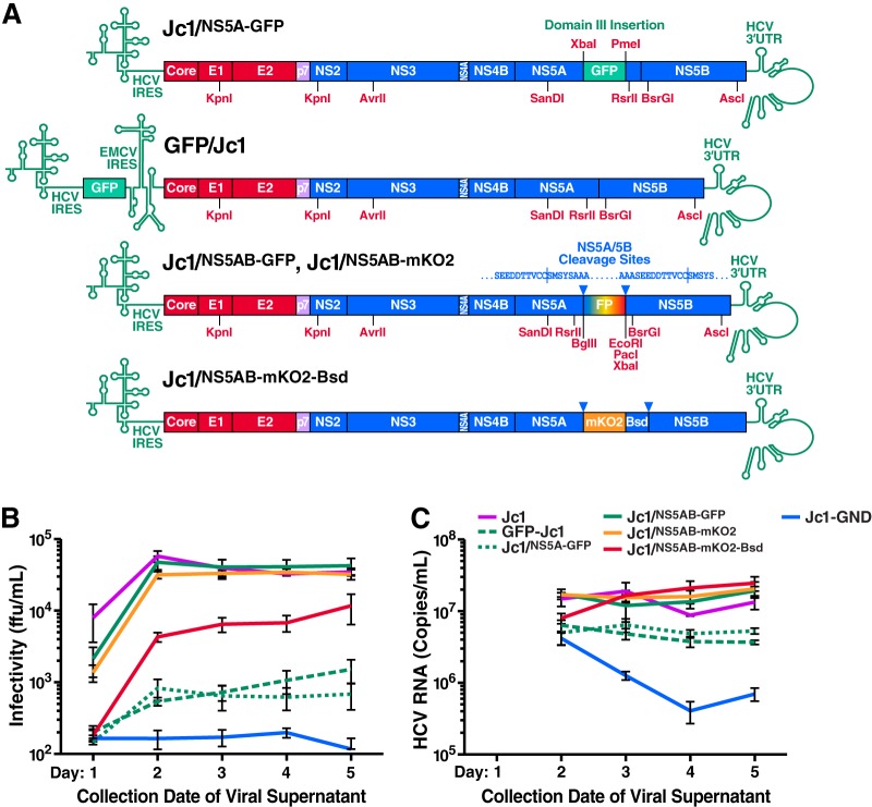
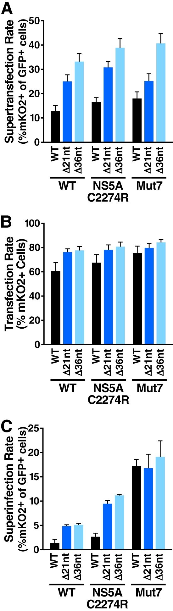
Cells that are productively infected by hepatitis C virus (HCV) are refractory to a second infection by HCV via a block in viral replication known as superinfection exclusion. The block occurs at a postentry step and likely involves translation or replication of the secondary viral RNA, but the mechanism is largely unknown. To characterize HCV superinfection exclusion, we selected for an HCV variant that could overcome the block. We produced a high-titer HC-J6/JFH1 (Jc1) viral genome with a fluorescent reporter inserted between NS5A and NS5B and used it to infect Huh7.5 cells containing a Jc1 replicon. With multiple passages of these infected cells, we isolated an HCV variant that can superinfect cells at high levels. Notably, the superinfectious virus rapidly cleared the primary replicon from superinfected cells. Viral competition experiments, using a novel strategy of sequence-barcoding viral strains, as well as superinfection of replicon cells demonstrated that mutations in E1, p7, NS5A, and the poly(U/UC) tract of the 3' untranslated region were important for superinfection. Furthermore, these mutations dramatically increased the infectivity of the virus in naive cells. Interestingly, viruses with a shorter poly(U/UC) and an NS5A domain II mutation were most effective in overcoming the postentry block. Neither of these changes affected viral RNA translation, indicating that the major barrier to postentry exclusion occurs at viral RNA replication. The evolution of the ability to superinfect after less than a month in culture and the concomitant exclusion of the primary replicon suggest that superinfection exclusion dramatically affects viral fitness and dynamics in vivo.


Figures

Fig 1
Fig 1
Construction of highly infectious reporter HCV strains. (A) Schematic diagram of HCV reporter strains. FP represents the fluorophore tag (GFP or mKO2) and Bsd represents the blasticidin resistance gene. (B and C) Huh7.5 cells were transfected with RNA transcripts of the given strains, including untagged Jc1. Supernatants were collected from days 1 to 5 after transfection and clarified by filtration. The nonreplicating Jc1-GND (polymerase defective, NS5B GDD→GND mutation) was used as a transfection control. (B) Titers of infectious HCV particles after transfection. Virus titers were obtained by limiting dilution assay. Note the higher infectious titers in the Jc1/NS5AB reporter strains relative to previously described reporter strains. (C) Release of HCV RNA after transfection. RNA was isolated from supernatants after transfection and HCV RNA was quantified by real-time RT-PCR. Note the higher viral RNA levels in the Jc1/NS5AB reporter strains relative to previously described reporter strains. n = 3 independent viral preparations and quantifications; error bars indicate the standard errors of the mean (SEM).
Fig 2
Fig 2
Isolation of a superinfectious HCV strain. (A) Schematic diagram of the Jc1/ΔE1E2NS5A-GFP replicon construct. (B) Diagram of the strategy used to isolate the superinfectious Jc1/NS5AB-mKO2-Bsd reporter strain. Briefly, a 1:1 mixture of HCV-naive EBFP2-tagged cells (H2B-EBFP2) and polyclonal Jc1/ΔE1E2NS5A-GFP replicon-containing cells were transfected with Jc1/NS5AB-mKO2-Bsd RNA. Three days later, these cells were labeled with FeOLabel magnetic beads and mixed with unlabeled H2B-EBFP2 and Jc1/ΔE1E2NS5A-GFP cells to allow the secondary Jc1/NS5AB-mKO2-Bsd virus to spread into the unlabeled cells. The magnetically labeled cells were removed 3 days later, and the Jc1/NS5AB-mKO2-Bsd-infected culture was continuously passaged. (C) Emergence of a superinfectious Jc1/NS5AB-mKO2-Bsd strain during continuous passage. The blue line indicates the percentage of H2B-EBFP2 “naive” cells determined by flow cytometry; these cells were quickly eliminated from the culture, likely by the Jc1/NS5AB-mKO2-Bsd virus. The orange line indicates the percentage of Jc1/ΔE1E2NS5A-GFP replicon-containing cells superinfected with Jc1/NS5AB-mKO2-Bsd. Note that emergence of the superinfectious strain at ∼27 days after separation correlates with elimination of the Jc1/ΔE1E2NS5A-GFP replicon (green line) from the culture. (D) Serial passage of viral supernatants over replicon-containing cultures to further select for a superinfectious phenotype. (E) Viral supernatants passaged for 10 rounds display high superinfection rates and the ability to exclude the primary replicon. WT or adapted (10 rounds of passage over replicon cells) Jc1/NS5AB-mKO2-Bsd viruses were used to superinfect polyclonal Jc1/ΔE1E2NS5A-GFP replicon-containing cells. The percentage of superinfected replicon+ cells and the total percentage of replicon+ cells were measured by flow cytometry at the given time points. n = 3 independent experiments; error bars indicate the SEM.
Fig 3
Fig 3
Contribution of identified mutations to the superinfectious phenotype. (A) Schematic diagram of the genotype 1b Con1 SGR/NS5A-GFP replicon. NeoR indicates the neomycin resistance gene. (B) Recapitulation of the superinfectious phenotype with seven mutations. Mutations were introduced into the WT Jc1/NS5AB-mKO2-Bsd construct, and viral supernatants were produced by transfection of these variants into Huh7.5 cells. Supernatants were normalized to the quantity of HCV core (determined by ELISA) and Jc1/ΔE1E2NS5A-GFP replicon-containing cells were superinfected. The percent superinfection was assessed by flow cytometry 3 days later. To ensure that the superinfectious phenotype was not limited to a particular Jc1/ΔE1E2NS5A-GFP replicon cell line, superinfections were performed in polyclonal and monoclonal replicon cells (replicon cell lines were isolated separately). Note that the NS5A D2437N mutation had a detrimental effect on superinfectivity and was thus excluded from the majority of variants analyzed. (C) Superinfectivity correlates with a higher degree of infectivity in naive cells. The same viruses used in panel A were used to infect naive Huh7.5 cells. (D) Superinfection is reduced in a genotype 1b replicon cell line. Polyclonal Jc1/ΔE1E2NS5A-GFP and Con1 SGR/NS5A-GFP replicon cells were superinfected with the given viral variants. (E) Both NS3 A1564T and NS4A H1691L increase superinfectivity alone but not when the mutations were combined. n = 4 independent viral preparations and infections; error bars indicate the SEM.
Fig 4
Fig 4
Superinfection spread in replicon cells demonstrates the importance of E1 A335S, p7 V756A, and NS5A C2274R mutations. Jc1/ΔE1E2NS5A-GFP monoclonal replicon-containing cells were superinfected at a multiplicity of infection of 0.1 (7.5 × 10⁴ FFU/ml), and cultures were continuously passaged for 22 days-postinfection. The superinfection rate (%mKO2+ of GFP+ cells) was analyzed at various time points by flow cytometry. Note that the superinfectious phenotype is still present even when viral input is normalized by infectious titer rather than HCV core; a long period of viral spread, however, is required to observe this phenotype. The data are representative of three independent viral preparations and experiments.
Fig 5
Fig 5
The highly infectious and superinfectious phenotype of the Mut7 and Mut6 viruses is not limited to the Jc1/NS5AB-mKO2-Bsd reporter strain. (A) Infectivity of supernatants from Mut7 and Mut6 Jc1-transfected cultures is ∼10-fold higher than WT Jc1-transfected cultures. Untagged Jc1 (WT, Mut7, or Mut6) viral supernatants produced by transfection of Huh7.5 cells were assessed for infectivity using the limiting dilution assay on naive Huh7.5 cells. (B) Exclusion of the Jc1/ΔE1E2NS5A-GFP primary replicon is enhanced in Mut7- and Mut6-superinfected cultures. Polyclonal Jc1/ΔE1E2NS5A-GFP replicon-containing cells were superinfected with untagged Jc1 (WT, Mut7, or Mut6) or the adapted superinfecting strain (rounds 10, 11, and 13) viral supernatants. Exclusion of the primary replicon was assessed by flow cytometry. (C) Exclusion of the Jc1/ΔE1E2NS5A-FLuc-Bsd primary replicon is enhanced in Mut7- and Mut6-superinfected cultures. Polyclonal 7.5-RLuc Jc1/ΔE1E2NS5A-FLuc-Bsd cells were superinfected with untagged Jc1 (WT, Mut7, or Mut6). 6 days postinfection, cells were analyzed for FLuc (replicon-derived) and RLuc (control, lentivirus-derived) luminescence. Note the greater loss of the primary replicon in the Mut7 Jc1 and Mut6 Jc1-superinfected cultures compared to WT Jc1-superinfected or nonsuperinfected cultures. n = 3 independent viral preparations and infections; error bars indicate ± the SEM. *, P < 0.05; ***, P < 0.001; ****, P < 0.0001 (one-way ANOVA or analysis of covariance).
Fig 6
Fig 6
Overview of the viral barcode cloning strategy. Viral RNA was isolated and subjected to one-step RT-PCR using the primers NS5ABmcs_1 and Jc1_Amplicon_15R. A nested PCR was performed using the primers Barcode_LIC_Fw_5 and Barcode_LIC_Rev_5 or the primers Barcode_LIC_Fw_3 and Barcode_LIC_Rev_3. Complementary regions on the 5′ ends of these primers caused the PCR products to anneal at these regions during later cycles; the resulting overlap extension led to the fusion of PCR products into long concatemers. Concatemers were purified by gel extraction and cloned into pCR Blunt II Topo. Colony PCR was then performed, followed by sequencing.
Fig 7
Fig 7
Competition between multiple viral variants shows that p7 V756A, NS5A C2274R, and Δ 3′UTR mutations are important in the superinfectious phenotype. Jc1/Barcode viral strains were introduced into naive Huh7.5 cells by transfection or infection. RNA was obtained from the transfection or infection input or from cell-free supernatants at the given time points posttransfection or postinfection. The relative prevalence of each Jc1/Barcode strain was determined by the barcode cloning strategy shown in Fig. 6. (A) WT Jc1 and strains lacking the p7 V756A, NS5A C2274R, and Δ 3′ UTR mutations are excluded from cotransfected cultures. A total of 1.11 μg of each Jc1/NS5AB-Barcode viral RNA was cotransfected into Huh7.5 cells. (B and C) The pattern of viral exclusion is similar in coinfected cells, whether viral input is normalized by RNA or infectivity. (B) Viral competition after coinfection with viral stocks normalized by RNA input. Huh7.5 cells were coinfected with 1.53 × 10⁶ viral genome equivalents of each Jc1/NS5AB-Barcode viral stock/ml. (C) Viral competition after coinfection with viral stocks normalized by infectivity. Huh7.5 cells were coinfected with 1.01 × 10⁴ FFU of each Jc1/NS5AB-Barcode viral stock/ml. n = 3 independent transfections or infections; error bars indicate the SEM.
Fig 8
Fig 8
NS5A C2274R and Δ 3′UTR mutations increase the amount of viral protein. (A) Sequence alignment of poly(U/UC) sequences from mutant and WT viral variants. The sequence from the superinfecting virus reflects the 24-nt deletion found in all sequenced clones; the length of the poly(U) tract varied according to each clone. Nucleotides are numbered according to the Jc1/NS5AB-mKO2-Bsd RNA. (B) Representative flow cytometry plot indicating an increase in the mean fluorescence intensity of mKO2 in Huh7.5 cells infected with WT or NS5A C2274R Jc1/NS5AB-mKO2-Bsd. Cells were infected with viral stocks normalized by core input and analyzed by flow cytometry at 3 days postinfection. (C) Western blots indicating an increase in HCV core and mKO2-Bsd in Huh7.5 cells transfected with HCV Jc1/NS5AB-mKO2-Bsd strains carrying the NS5A C2274R and Δ 3′ UTR mutations. Cells were transfected with 10 μg of viral RNA, and lysates were obtained 3 days posttransfection.
Fig 9
Fig 9
NS5A C2274R and Δ 3′UTR mutations increase “supertransfection” in replicon-containing cells. (A) “Supertransfection” is greatly enhanced by poly(U/UC) deletions and slightly enhanced by the NS5A C2274R mutation. Jc1/ΔE1E2NS5A-GFP polyclonal replicon cells were transfected with WT and mutant Jc1/NS5AB-mKO2-Bsd transcripts. The “supertransfection” rate was assessed by flow cytometry as the percentage of replicon-positive cells (GFP+) that were also positive for the secondary Jc1/NS5AB-mKO2-Bsd virus (mKO2+) 2 days posttransfection. n = 4 independent transfections. (B) Transfection rates in naive cells are also enhanced by the poly(U/UC) deletion and NS5A C2274R mutation. Naive Huh7.5 cells were transfected with WT and mutant Jc1/NS5AB-mKO2-Bsd transcripts and transfection rates (% mKO2+) were assessed by flow cytometry 2 days later. n = 4 independent transfections. (C) Superinfection rates are also enhanced by the poly(U/UC) deletion and NS5A C2274R mutation. Jc1/ΔE1E2NS5A-GFP polyclonal replicon cells were infected with WT and mutant Jc1/NS5AB-mKO2-Bsd viral supernatants (normalized to the amount of HCV core per infection). Superinfection rates were assessed by flow cytometry 3 days later. n = 3 independent viral preparations and infections; error bars indicate the SEM.
Fig 10
Fig 10
Viral RNA translation is not enhanced by the NS5A C2274R and Δ 3′UTR mutations. (A) Deletions of the poly(U/UC) tract do not enhance HCV IRES-mediated translation. Jc1/ΔE1E2NS5A-GFP polyclonal replicon cells and IFN-α replicon-cured cells were transfected with HCV IRES-dependent firefly luciferase (FLuc) transcripts containing WT and mutant HCV 3′UTRs, as well as a capped Renilla luciferase (RLuc) transcript as a transfection control. Core+262 refers to a fusion of 262 nt of HC-J6 core sequence to FLuc to enhance HCV IRES-mediated translation. The luciferase activity was assessed at 2 h posttransfection, and the ratio of FLuc/RLuc activity was normalized to the WT 3′UTR construct in replicon-cured cells. n = 3 independent transfections. (B) Histogram of normalized FLuc/RLuc ratios showing that NS5A C2274R and Δ 3′UTR mutations do not enhance translation of replication-incompetent viral RNAs. Cells were transfected as in panel A with Jc1/NS5AB-FLuc-GND transcripts (polymerase defective, NS5B GDD→GND mutation) containing NS5A C2274R or Δ 3′UTR mutations. Arrowheads indicate NS5A/5B protease cleavage sites. n = 3 independent transfections of two replicates each; error bars indicate the SEM.

Similar articles

Cited by

References

    1. Michel N, Allespach I, Venzke S, Fackler OT, Keppler OT. 2005. The Nef protein of human immunodeficiency virus establishes superinfection immunity by a dual strategy to downregulate cell surface CCR5 and CD4. Curr. Biol. 15:714–723 - PubMed
    1. Adams RH, Brown DT. 1985. BHK cells expressing Sindbis virus-induced homologous interference allow the translation of nonstructural genes of superinfecting virus. J. Virol. 54:351–357 - PMC - PubMed
    1. Walters KA, Joyce MA, Addison WR, Fischer KP, Tyrrell DL. 2004. Superinfection exclusion in duck hepatitis B virus infection is mediated by the large surface antigen. J. Virol. 78:7925–7937 - PMC - PubMed
    1. Folimonova SY. 2012. Superinfection exclusion is an active virus-controlled function that requires a specific viral protein. J. Virol. 86:5554–5561 - PMC - PubMed
    1. Schaller T, Appel N, Koutsoudakis G, Kallis S, Lohmann V, Pietschmann T, Bartenschlager R. 2007. Analysis of hepatitis C virus superinfection exclusion by using novel fluorochrome gene-tagged viral genomes. J. Virol. 81:4591–4603 - PMC - PubMed

Publication types