. 2013:2013:678473.
doi: 10.1155/2013/678473. Epub 2013 Sep 5.

Lack of HXK2 induces localization of active Ras in mitochondria and triggers apoptosis in the yeast Saccharomyces cerevisiae

Affiliations

Lack of HXK2 induces localization of active Ras in mitochondria and triggers apoptosis in the yeast Saccharomyces cerevisiae

Loredana Amigoni et al. Oxid Med Cell Longev. 2013.

Abstract

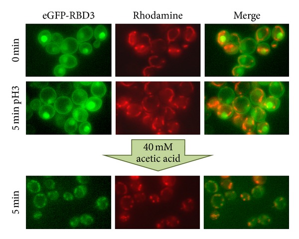
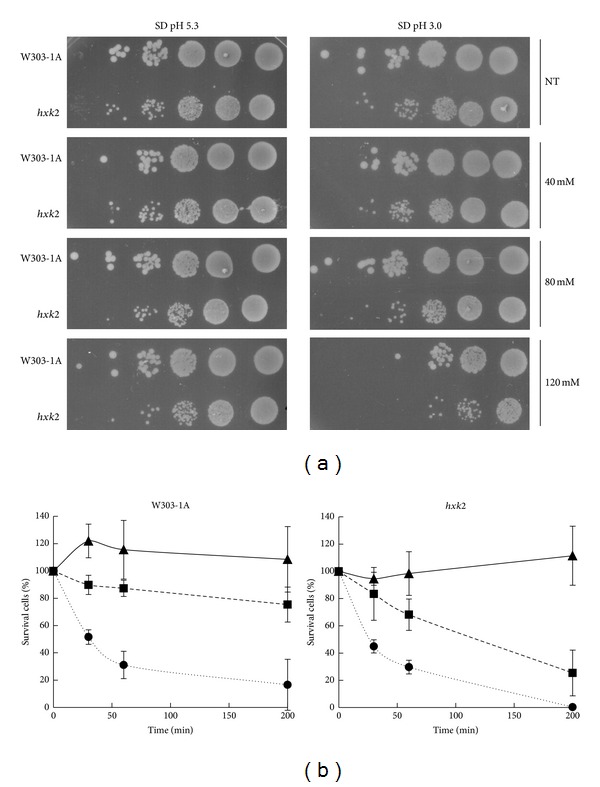
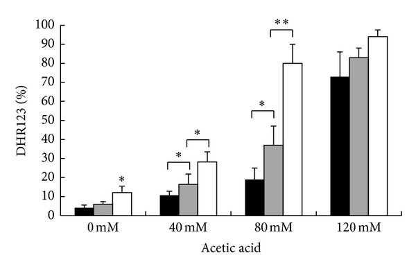
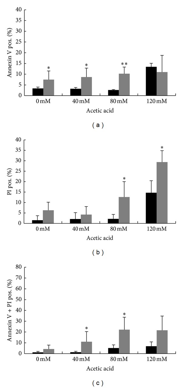
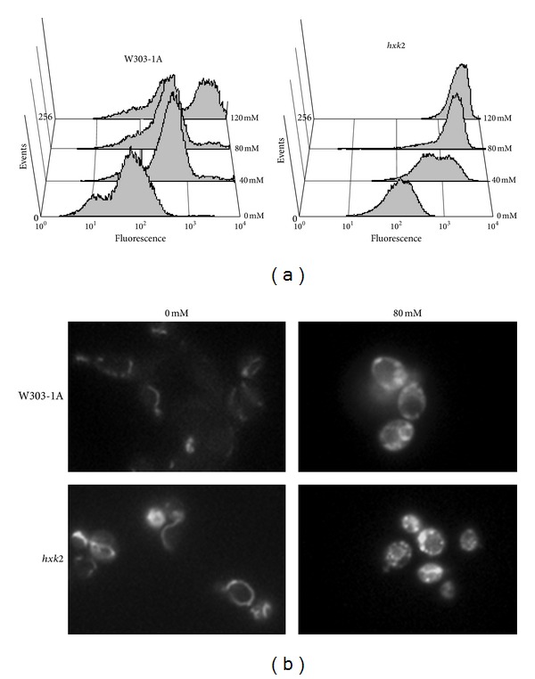
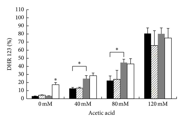
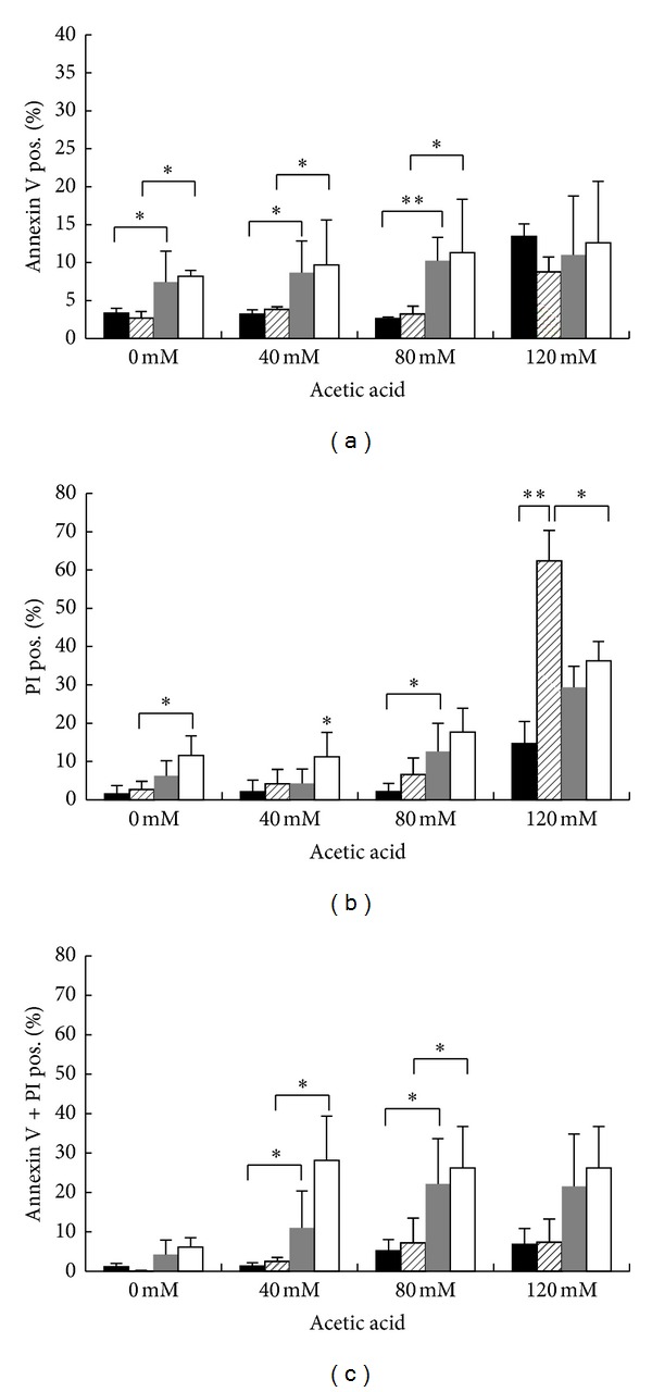
We recently showed that activated Ras proteins are localized to the plasma membrane and in the nucleus in wild-type cells growing exponentially on glucose, while in the hxk2Δ strain they accumulated mainly in mitochondria. An aberrant accumulation of activated Ras in these organelles was previously reported and correlated to mitochondrial dysfunction, accumulation of ROS, and cell death. Here we show that addition of acetic acid to wild-type cells results in a rapid recruitment of Ras-GTP from the nucleus and the plasma membrane to the mitochondria, providing a further proof that Ras proteins might be involved in programmed cell death. Moreover, we show that Hxk2 protects against apoptosis in S. cerevisiae. In particular, cells lacking HXK2 and showing a constitutive accumulation of activated Ras at the mitochondria are more sensitive to acetic-acid-induced programmed cell death compared to the wild type strain. Indeed, deletion of HXK2 causes an increase of apoptotic cells with several morphological and biochemical changes that are typical of apoptosis, including DNA fragmentation, externalization of phosphatidylserine, and ROS production. Finally, our results suggest that apoptosis induced by lack of Hxk2 may not require the activation of Yca1, the metacaspase homologue identified in yeast.


Figures

Figure 1
Figure 1
Localization of active Ras in the W303-1A wild-type strain after addition of 40 mM acetic acid. Cells transformed with YEpeGFP-RBD3 were grown in 2% glucose medium at 30°C until exponential phase, collected by centrifugation, and resuspended in medium adjusted to pH 3.0. Cells were then photographed with a Nikon fluorescence microscope, before and after addition of 40 Mm acetic acid. Colocalization of eGFP fluorescence and the red-fluorescent Rhodamine B hexyl ester is clearly visible five minutes after addition of the apoptotic stimulus.
Figure 2
Figure 2
hxk2Δ cells exhibit inhibition of cell growth and hypersensitivity to acetic acid. (a) W303-1A and hxk2Δ cells were harvested and resuspended (1-2 × 107 cells/mL) in SD medium adjusted either at pH 5.3 or at pH 3.0 (set with HCl) in the absence (NT) or in the presence of 40, 80, or 120 mM acetic acid. Cells were incubated for 200 min at 30°C with shaking (160 rpm). After treatment, cells were harvested and resuspended at the same concentration (108 cells/mL) in milliQ water. 5 microliter from a concentrated suspension and from 10-fold dilutions of each culture was spotted onto YEPD plates and incubated at 30°C for 3 days. (b) Cell survival of W303-1A and hxk2Δ strains. Cell viability of W303-1A and hxk2Δ untreated cells (▲) or treated with 80 mM (■) and 120 mM () acetic acid was analyzed at indicated times by measuring colony-forming units (cfu) after 2 days of growth at 30°C. Cell survival (100%) corresponds to the cfu at time zero. The means of 4 independent experiments with standard deviations are reported.
Figure 3
Figure 3
ROS accumulation in W303-1A, hxk2Δ, and hxk2Δ cells expressing the activated Ras2 Val19 allele after treatment with acetic acid. W303-1A (black bars), hxk2Δ (gray bars), and hxk2Δ cells expressing the activated Ras2Val19 allele (white bars) exponentially growing cells were treated with different concentrations (40–80–120 mM) of acetic acid for 200 minutes at 30°C. ROS accumulation was assayed using the dye dihydrorhodamine 123 (DHR123) by flow cytometry. The means of 3 independent experiments with standard deviations are reported. Student's t-test *P < 0.05 and **P < 0.01.
Figure 4
Figure 4
The occurrence of acetic-acid-induced cell death in hxk2Δ cells is characterized by markers of both apoptosis and necrosis. Assessment of cell death by FITC-coupled annexin V and PI staining. W303-1A (black bars) and hxk2Δ (gray bars) exponentially growing cells were treated with different concentrations (40–80–120 mM) of acetic acid for 200 minutes at 30°C, before being processed for determination of phosphatidylserine externalization and membrane integrity by flow cytometry. 30000 events have been evaluated. The means of 3 independent experiments with standard deviations are reported. Student's t-test *P < 0.05 and **P < 0.01.
Figure 5
Figure 5
Mitochondrial membrane potential and morphology in W303-1A and hxk2Δ strains upon acetic acid treatment. W303-1A and hxk2Δ exponentially growing cells were treated with different concentrations (40–80–120 mM) of acetic acid for 200 minutes at 30°C. DiOC6 uptake was assessed using both flow cytometry (a) and fluorescence microscopy (b). Photomicrographs illustrate the alteration of the tubular mitochondrial network to clustered mitochondrial morphology, particularly exacerbated in hxk2Δ cells after acetic acid treatment.
Figure 6
Figure 6
Effect of YCA1 deletion on ROS accumulation after treatment with acetic acid. W303-1A (black bars), yca1Δ (black hatched bars), hxk2Δ (gray bars), and hxk2Δ-yca1Δ (white bars) exponentially growing cells were treated with different concentrations (40–80–120 mM) of acetic acid for 200 minutes at 30°C. ROS accumulation was assayed by flow cytometry using the dye dihydrorhodamine 123 (DHR123). Student's t-test *P < 0.05.
Figure 7
Figure 7
Effect of YCA1 deletion on cell death after treatment with acetic acid. W303-1A (black bars), yca1Δ (black hatched bars), hxk2Δ (gray bars) and hxk2Δ-yca1Δ (white bars) exponentially growing cells were treated with different concentrations (40–80–120 mM) of acetic acid for 200 minutes at 30°C. Cell death was assessed by flow cytometry using FITC-coupled annexin V and PI co-staining to determinate the externalization of phosphatidylserine and the membrane integrity. The means of 3 independent experiments with standard deviations are reported. Student's t-test *P < 0.05 and **P < 0.01.

References

    1. Jones S, Vignais M-L, Broach JR. The CDC25 protein of Saccharomyces cerevisiae promotes exchange of guanine nucleotides bound to Ras. Molecular and Cellular Biology. 1991;11(5):2641–2646. - PMC - PubMed
    1. Rudoni S, Colombo S, Coccetti P, Martegani E. Role of guanine nucleotides in the regulation of the Ras/cAMP pathway in Saccharomyces cerevisiae . Biochimica et Biophysica Acta. 2001;1538(2-3):181–189. - PubMed
    1. Tanaka K, Nakafuku M, Satoh T, et al. S. cerevisiae genes IRA1 and IRA2 encode proteins that may be functionally equivalent to mammalian ras GTPase activating protein. Cell. 1990;60(5):803–807. - PubMed
    1. Zaman S, Lippman SI, Zhao X, Broach JR. How Saccharomyces responds to nutrients. Annual Review of Genetics. 2008;42:27–81. - PubMed
    1. Thevelein JM, De Winde JH. Novel sensing mechanisms and targets for the cAMP-protein kinase A pathway in the yeast Saccharomyces cerevisiae . Molecular Microbiology. 1999;33(5):904–918. - PubMed

Publication types

MeSH terms

LinkOut - more resources