

Evidence and recommendations for imaging liver fat in children, based on systematic review
- PMID: 24090729
- PMCID: PMC3969892
- DOI: 10.1016/j.cgh.2013.09.050
Evidence and recommendations for imaging liver fat in children, based on systematic review
Abstract
Background & aims: Fatty liver is a common problem in children and increases their risk for cirrhosis, diabetes, and cardiovascular disease. Liver biopsy is the clinical standard for diagnosing and grading fatty liver. However, noninvasive imaging modalities are needed to assess liver fat in children. We performed a systematic review of studies that evaluated imaging liver fat in children.
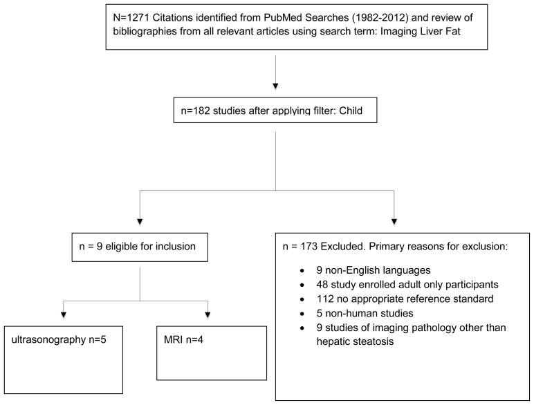
Methods: We searched PubMed for original research articles in peer-reviewed journals from January 1, 1982, through December 31, 2012, using the key words "imaging liver fat." Studies included those in English, and those performed in children from birth to 18 years of age. To be eligible for inclusion, studies were required to measure hepatic steatosis via an imaging modality and a quantitative comparator as the reference standard.
Results: We analyzed 9 studies comprising 610 children; 4 studies assessed ultrasonography and 5 studies assessed magnetic resonance imaging (MRI). Ultrasonography was used in the diagnosis of fatty liver with positive predictive values of 47% to 62%. There was not a consistent relationship between ultrasound steatosis score and the reference measurement of hepatic steatosis. Liver fat as measurements by MRI or by spectroscopy varied with the methodologies used. Liver fat measurements by MRI correlated with results from histologic analyses, but sample size did not allow for an assessment of diagnostic accuracy.
Conclusions: Available evidence does not support the use of ultrasonography for the diagnosis or grading of fatty liver in children. Although MRI is a promising approach, the data are insufficient to make evidence-based recommendations regarding its use in children for the assessment of hepatic steatosis.
Keywords: Evidence-Based Medicine; Liver Imaging; NAFLD.
Copyright © 2014 AGA Institute. Published by Elsevier Inc. All rights reserved.
Conflict of interest statement
Figures
References
-
- Schwimmer JB, et al. Prevalence of fatty liver in children and adolescents. Pediatrics. 2006;118(4):1388–93. - PubMed
-
- Lee SS, et al. Non-invasive assessment of hepatic steatosis: prospective comparison of the accuracy of imaging examinations. J Hepatol. 2010;52(4):579–85. - PubMed
-
- Mennesson N, et al. Liver steatosis quantification using magnetic resonance imaging: a prospective comparative study with liver biopsy. J Comput Assist Tomogr. 2009;33(5):672–7. - PubMed
Publication types
MeSH terms
Grants and funding
LinkOut - more resources
Full Text Sources
Other Literature Sources
Medical
