. 2014 Jan;1838(1 Pt B):185-92.
doi: 10.1016/j.bbamem.2013.09.014. Epub 2013 Sep 30.

Interaction of Plasmodium falciparum knob-associated histidine-rich protein (KAHRP) with erythrocyte ankyrin R is required for its attachment to the erythrocyte membrane

Affiliations

Interaction of Plasmodium falciparum knob-associated histidine-rich protein (KAHRP) with erythrocyte ankyrin R is required for its attachment to the erythrocyte membrane

Haibo Weng et al. Biochim Biophys Acta. 2014 Jan.

Abstract

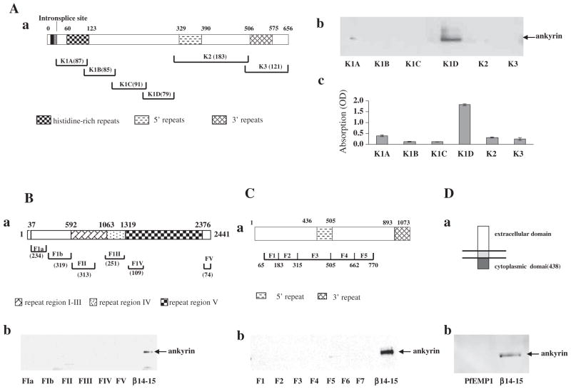
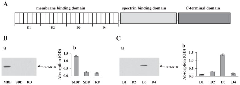
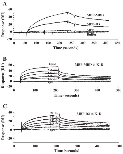
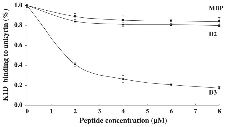
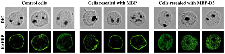
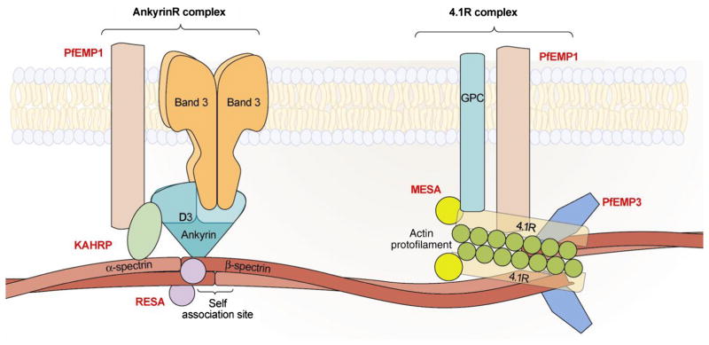
The malaria parasite Plasmodium falciparum exports a large number of proteins into the erythrocyte cytoplasm during the asexual intraerythrocytic stage of its life cycle. A subset of these proteins interacts with erythrocyte membrane skeletal proteins and grossly alters the structure and function of the membrane. Several of the exported proteins, such as PfEMP1, PfEMP3, RESA and KAHRP, interact with the preponderant erythrocyte skeleton protein, spectrin. Here we have searched for possible interaction of these four malaria proteins with another major erythrocyte skeleton protein, ankyrin R. We have shown that KAHRP, but none of the other three, binds to ankyrin R. We have mapped the binding site for ankyrin R to a 79-residue segment of the KAHRP sequence, and the reciprocal binding site for KAHRP in ankyrin R to a subdomain (D3) of the 89kDa ankyrin R membrane-binding domain. Interaction of intact ankyrin R with KAHRP was inhibited by the free D3 subdomain. When, moreover, red cells loaded with the soluble D3 subdomain were infected with P. falciparum, KAHRP secreted by the intraerythrocytic parasite no longer migrated to the host cell membrane, but remained diffusely distributed throughout the cytosol. Our findings suggest a potentially important role for interaction of KAHRP with red cell membrane skeleton in promoting the adhesion of malaria-infected red cells to endothelial surfaces, a central element in the pathophysiology of malaria.

Keywords: Ankyrin R; Cytoskeletal proteins; KAHRP; Malaria; Red cell.


Figures

Fig. 1
Fig. 1
Association of malaria proteins with ankyrin R. (A) Binding of ankyrin R to KAHRP fragments. Schematic representation of KAHRP (a). The binding between ankyrin R and KAHRP fragments was assessed by pull-down (b) and ELISA assay (c). Note that ankyrin R bound to only the K1D fragment of the KAHRP. (B) Binding of ankyrin R to PfEMP3 fragments. Schematic representation of PfEMP3 (a). The binding between ankyrin R and PfEMP3 fragments was assessed by pull-down (b). Repeats 14–15 fragment of β-spectrin was used as a positive control for ankyrin R binding. (C) Binding of ankyrin R to RESA fragments. Schematic representation of RESA (a). The binding between ankyrin R and RESA fragments was assessed by pull-down (b). Repeats 14–15 fragment of β-spectrin was used as a positive control for ankyrin R binding. (D) Binding of ankyrin R to the cytoplasmic domain of PfEMP1. Schematic representation of PfEMP1 (a). The binding between ankyrin R and cytoplasmic domain of PfEMP1 was assessed by pull-down (b). Repeats 14–15 fragment of β-spectrin was used as a positive control for ankyrin R binding. Amino acid residue numbers and the lengths of fragments are indicated.
Fig. 2
Fig. 2
Binding site of KAHRP in ankyrin R. (A) Schematic representation of ankyrin R functional domains. (B) Binding of K1D to functional domains of ankyrin R. The binding of GST-tagged K1D fragment to MBP-tagged ankyrin R domains was assessed by MBP pull-down assay (left panel) and ELISA (right). Note that K1D bound to only the MBD of ankyrin R. (C) Binding of K1D fragment to subdomains of the MBD domain of ankyrin R. The binding of GST-tagged K1D fragment to sub-domains of MBD was assessed by MBP pull-down assay (a) and by ELISA assay (b). Note that K1D bound to only the D3 region of the MBD domain of ankyrin R.
Fig. 3
Fig. 3
Kinetic analysis of interaction between K1D and ankyrin R polypeptides by surface plasmon resonance assay. A: GST-tagged K1D fragment was immobilized onto a CM5 sensor chip. MBP, MBP-MBD or MBP-D3 at a concentration of 1 μM was injected at 20 μl/min over the chip surface in a BIAcore 3000 instrument. B: MBP-MBD at different concentrations as indicated was injected at 20 μl/min over the surface in a BIAcore 3000 instrument. The figure shows dose–response curves of MBP-MBD binding to K1D. C: MBP-D3 at different concentrations as indicated was injected at 20 μl/min over the surface in a BIAcore 3000 instrument. The figure shows dose–response curves of MBP-D3 binding to K1D.
Fig. 4
Fig. 4
Inhibition of binding of K1D to ankyrin R by the D3 fragment of ankyrin R. K1D was incubated with increasing concentrations of D3 or MBP before addition to immobilized ankyrin R in the ELISA wells. K1D binding to ankyrin R is inhibited by D3.
Fig. 5
Fig. 5
Localization and distribution of KAHRP in erythrocytes infected with P. falciparum. Erythrocytes were resealed with no added proteins (A), with 40 μM MBP (B), or with 40 μM MBP-D3 (C). The resealed erythrocytes were infected with P. falciparum and cultured for 48 h. The localization of KAHRP was detected by immunostaining with an anti-KAHRP antibody (green color). The stained cells were imaged by confocal microscopy (100× objection).
Fig. 6
Fig. 6
Schematic representation of interactions between malaria proteins and host skeleton proteins in malaria infected erythrocytes. At the ankyrin R-based macromolecular complex, which is near the center of the tetramer (dimer–dimer interaction site), PfEMP1 binds to KAHRP which in turn binds to both ankyrin R and α-spectrin. RESA binds to repeat 16 of β-spectrin. At the 4.1R-based macromolecular complex, PfEMP1 is anchored to the junctional complex through its direct binding to spectrin, actin and 4.1R. PfEMP3 also directly binds to spectrin, actin and 4.1R. MESA binds to the 30 kDa membrane domain of 4.1R.

References

    1. Hiller NL, Bhattacharjee S, van Ooij C, Liolios K, Harrison T, et al. A host-targeting signal in virulence proteins reveals a secretome in malarial infection. Science. 2004;306:1934–1937. - PubMed
    1. Marti M, Baum J, Rug M, Tilley L, Cowman AF. Signal-mediated export of proteins from the malaria parasite to the host erythrocyte. J Cell Biol. 2005;171:587–592. - PMC - PubMed
    1. Sargeant TJ, Marti M, Caler E, Carlton JM, Simpson K, et al. Lineage-specific expansion of proteins exported to erythrocytes in malaria parasites. Genome Biol. 2006;7:R12. - PMC - PubMed
    1. Oh SS, Chishti AH, Palek J, Liu SC. Erythrocyte membrane alterations in Plasmodium falciparum malaria sequestration. Curr Opin Hematol. 1997;4:148–154. - PubMed
    1. Maier AG, Cooke BM, Cowman AF, Tilley L. Malaria parasite proteins that remodel the host erythrocyte. Nat Rev Microbiol. 2009;7:341–354. - PubMed

Publication types

MeSH terms

Substances

LinkOut - more resources