. 2013 Sep 30;26(6):629-44.
doi: 10.1016/j.devcel.2013.07.019.

Integration of left-right Pitx2 transcription and Wnt signaling drives asymmetric gut morphogenesis via Daam2

Affiliations

Integration of left-right Pitx2 transcription and Wnt signaling drives asymmetric gut morphogenesis via Daam2

Ian C Welsh et al. Dev Cell. .

Abstract

A critical aspect of gut morphogenesis is initiation of a leftward tilt, and failure to do so leads to gut malrotation and volvulus. The direction of tilt is specified by asymmetric cell behaviors within the dorsal mesentery (DM), which suspends the gut tube, and is downstream of Pitx2, the key transcription factor responsible for the transfer of left-right (L-R) information from early gastrulation to morphogenesis. Although Pitx2 is a master regulator of L-R organ development, its cellular targets that drive asymmetric morphogenesis are not known. Using laser microdissection and targeted gene misexpression in the chicken DM, we show that Pitx2-specific effectors mediate Wnt signaling to activate the formin Daam2, a key Wnt effector and itself a Pitx2 target, linking actin dynamics to cadherin-based junctions to ultimately generate asymmetric cell behaviors. Our work highlights how integration of two conserved cascades may be the ultimate force through which Pitx2 sculpts L-R organs.


Figures

FIG. 1
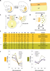
FIG. 1. DM and laser capture microdissection (LCM)
A Gut tube (GT, yellow; chicken HH20; mouse E10.0) undergoes counterclockwise rotation at HH21. B Gut tube is suspended by DM (orange) along the D-V axis. C Pitx2-driven deformation of DM at HH21 along the L-R axis initiates gut rotation. D LCM of the DM. E Selected microarray expression values from epithelium (E) and mesenchyme (M) of the Left (L) or Right (R) chicken DM (at HH21). Raw and normalized expression values are shown. Left genes (orange), right (green), and unbiased (yellow) are grouped. F Plot of normalized expression fold changes across the L-R axis.
FIG. 2
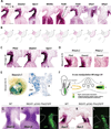
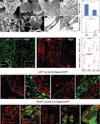
FIG. 2. Asymmetric organization of a Wnt signaling network across the L-R DM
A ISH, purple reveals positive (Daam2, Gpc3, Wnt5a, Fzd8, Fzd4) and negative (Sfrp1, and Sfrp2) Wnt pathway components in the DM and gut. B Gene expression schematic from A. Also see Fig. S1. C Left-sided Daam2 and absence of Daam2 in Pitx2−/− DM. D Left-sided Daam2 and Gpc3 prior to tilt. E Daam2tm1a(KOMP)Wtsi mice shows left-specific β-galactosidase activity. F Schematic of in ovo electroporation: DNA microinjected into the coelomic cavity (HH14) and electroporated to target the right splanchnic mesoderm (see IL). Arrow represents current-driven plasmid movement. G WT left-sided Daam2 expression. H pCAG-Pitx2 induces right-sided Daam2, marked by GFP in I. J WT left-sided Gpc3 expression. K pCAG-Pitx2 induces right-sided Gpc3, marked by GFP in L. Scale bars: A (100 µm); CIJKL (100 µm) D (50 µm) E (20 µm) See also Figure S1.
FIG. 3
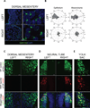
FIG. 3. Cellular behavior in the DM is polarized
A WT left and right DM stained with GM130 (golgi) and DAPI. B Cell orientation measured by the angle of the golgi with respect to the nucleus (X-axis: L-R, Y-axis: D-V, Radial-axis: number of cells per bin). Polarized left mesenchyme is oriented to the left (L: p<0.009; R: p<0.6). For L vs. R epithelium: p value < 0.0001). C Electroporation of pTOP-nRFP (red) into (C) the left and right DM; (D) neural tube; and (E) yolk sack (Also see Fig. S2). GFP identifies electroporated cells. Blue is DAPI. Scale bars: A (10 µm; insets are 5 µm); C (10µm).
FIG. 4
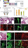
FIG. 4. Daam2 activation is required for mesenchymal condensation in the DM
A Wnt signal-dependent binding of Dishevelled to DAD activates Daam. B In ovo electroporations: N-Daam2 is targeted to the left, CA-Daam2 to the right (See also Fig. S4 for construct validation). C–H CA-Daam2 targeting of the right DM: C WT DM with left-specific condensation (yellow line) with asymmetric F-actin (red in F, Phalloidin); D CA-Daam2 induces condensation (green line, GFP coelectroporated in E) and (G) increased F-actin (GFP coelectroporated in H). I Right-sided Daam2 electroporation in the DM has no effect on condensation, marked by GFP in J. K–O’ N-Daam2 targeting of the left DM. K Loss of condensation (green boundary from L). White arrows in L show N-Daam2-electroporated cells separating from the left. M Control (pCAG-GFP) highlights tightly compacted cells compared to dispersed cells expressing N-Daam2 (N), which produce filopodia-like extensions (white arrows) and (O) exhibit decreased F-actin (red, Phalloidin). P Calculated mesenchymal cell densities of WT and electroporated tissue sections (mean ± SEM, all p-values <0.0155). Scale bars: C–H (50 µm); I–J (100 µm); K–L (50 µm); M–O’ (15µm). *, p<0.05. See also Figure S4–7.
FIG. 5
FIG. 5. Daam2 lengthens cell-cell junctions
A–G Scanning (A–C) and transmission (DG) EM show differences in left (ABDE) vs. right (ACFG) mesenchymal cell junction morphologies (yellow arrowheads). HI Cell-cell contacts on the left are increased in number (H, mean ± SEM) and length (I) vs. the right (p<0.05 & <0.01, respectively). (JK) Differences in cell junction organization highlighted by α-catenin (green, J) and N-cadherin staining (red, K, high magnifications inset); L-R boundary indicated (dotted line). LM Morphometric analysis of mean cell junction lengths in WT and electroporated tissues stained with α-catenin (top) and N-cadherin (bottom). Right-sided CA-Daam2 promotes lengthened junctions (p<0.0001) not statistically different from WT left cells (p>0.17), while left-sided N-Daam2 reduces junction length (p<0.0001) to resemble right-sided WT cells (p>0.59). N–O’ N-Daam2 (left) impairs adhesion and accumulation of both N-cadherin (red, NN’, note filopodia-like extensions, white arrows in N’) and α-catenin (red, OO’) to cell-cell contacts (GFP-marked electroporation boundary depicted). P–R CA-Daam2 (right) induces accumulation of both N-cadherin (red, PP’, yellow arrowheads) and α-catenin (red, Q–R). Scale bars: A (30µm top and 5µm bottom panels); EG (500nm); JK (10µm); NO’ (10µm); PP’ (5µm); QQ’ (10µm); R (5µm). Boxplots represent quartiles and median of the data, whiskers indicate extreme values. *, p<0.05, **, p<0.001.
FIG. 6
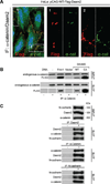
FIG. 6. Daam2 physically interacts with junctional proteins
A Flag-Daam2 (red) and endogenous α-catenin (green) co-localize at the cell surface in HeLa cells. Dotted rectangles (1 and 2) are magnified. B Overexpressed WT- and CA-Daam2, and positive control Fmn1 bind endogenous α-catenin. C Reciprocal IP experiments show Daam2 interacts with both α-catenin and N-cadherin (endognous, untransfected lysates). Scale bars: A (10µm).
FIG. 7
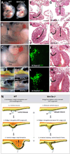
FIG. 7. Wnt5a−/− embryos fail to initiate the leftward tilt of the midgut
A Wnt5a−/− and WT embryos (E10.5, arrow marks DM). BC Higher magnification of A highlights arrested DM development in Wnt5a−/− embryo (C, dotted lines). D–E’ Dissection of embryos from A highlights DM (orange) and midgut (yellow). FH Normal leftward tilt (arrow) and loss of tilt in Wnt5a−/− embryo (GI). Dotted rectangles highlight arrested ventral outgrowth of the DM at the level of the midgut (F, WT; G, Wnt5a−/−) and defective morphology of the DM (H, WT; I, Wnt5a−/−) posterior to the midgut. J–M Chicken DM with N-Daam2 on the left (JL, GFP, electroporated). KM Coelomic cavity defects (black arrows) in N-Daam2 electroporated chick embryos are reminiscent of Wnt5a−/− embryos (I, black arrows). N Model for the role of Wnt5a in midgut loop formation: Ventral closure of the gut occurs at the initiation of midgut elongation and results in formation of the vitelline duct (vd). Delayed closure and arrested DM outgrowth in Wnt5a−/− forces the elongating midgut to branch rather than loop. Scale bars: B–E’ (500 µm); F (100 µm); GH (50 µm); I (100 µm); J–M (50 µm). See also Figure S5 and S1.
FIG. 8
FIG. 8. Model for L-R gut rotation
Transcriptional regulation of Wnt pathway genes by Pitx2 leads to Daam2 activation. Daam2 mediates adhesion at cell junctions by binding α-catenin and N-cadherin. Subsequent actin remodeling and lengthening of junctions cause left condensation. Wnt5a produced in the adjacent gut orients condensation relative to the D-V axis. Antagonized Wnt signaling causes right mesenchymal cells to remain dispersed.

Comment in

  • Your gut is right to turn left.
    Klezovitch O, Vasioukhin V. Klezovitch O, et al. Dev Cell. 2013 Sep 30;26(6):553-4. doi: 10.1016/j.devcel.2013.08.018. Dev Cell. 2013. PMID: 24091006 Free PMC article.

Similar articles

Cited by

References

    1. Aspenstrom P, Richnau N, Johansson AS. The diaphanous-related formin DAAM1 collaborates with the Rho GTPases RhoA and Cdc42, CIP4 and Src in regulating cell morphogenesis and actin dynamics. Exp Cell Res. 2006;312:2180–2194. - PubMed
    1. Baeg GH, Lin X, Khare N, Baumgartner S, Perrimon N. Heparan sulfate proteoglycans are critical for the organization of the extracellular distribution of Wingless. Development. 2001;128:87–94. - PubMed
    1. Barolo S. Transgenic Wnt/TCF pathway reporters: all you need is Lef? Oncogene. 2006;25:7505–7511. - PubMed
    1. Biechele TL, Adams AM, Moon RT. Transcription-based reporters of Wnt/beta-catenin signaling. Cold Spring Harbor protocols. 2009;2009 pdb prot5223. - PubMed
    1. Brent AE, Schweitzer R, Tabin CJ. A somitic compartment of tendon progenitors. Cell. 2003;113:235–248. - PubMed

Publication types

MeSH terms