. 2014 Apr;35(4):784-9.
doi: 10.3174/ajnr.A3744. Epub 2013 Oct 3.

Spontaneous lateral sphenoid cephaloceles: anatomic factors contributing to pathogenesis and proposed classification

Affiliations

Spontaneous lateral sphenoid cephaloceles: anatomic factors contributing to pathogenesis and proposed classification

F Settecase et al. AJNR Am J Neuroradiol. 2014 Apr.

Abstract

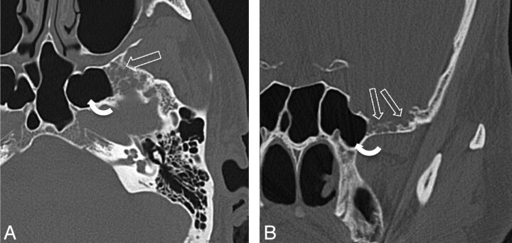
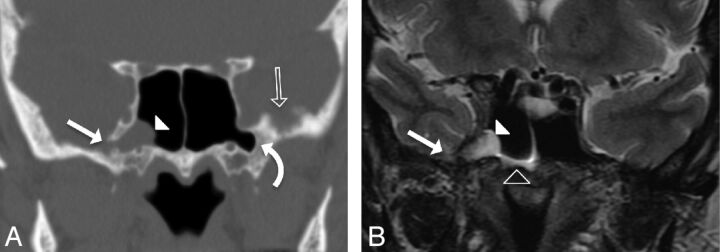
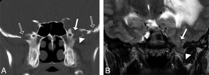
Spontaneous lateral sphenoid cephaloceles arise from bony defects in the lateral sphenoid, in the absence of predisposing factors such as trauma, surgery, mass, or congenital skull base malformation. We reviewed CT and MR imaging findings and clinical data of 26 patients with spontaneous lateral sphenoid cephaloceles to better understand anatomic contributions to pathogenesis, varying clinical and imaging manifestations, and descriptive terminology. Two types of spontaneous lateral sphenoid cephaloceles were identified. In 15 of 26 patients, a type 1 spontaneous lateral sphenoid cephalocele was noted, herniating into a pneumatized lateral recess of the sphenoid sinus, and typically presenting with CSF leak and/or headache. In 11 of 26 patients, a type 2 spontaneous lateral sphenoid cephalocele was noted, isolated to the greater sphenoid wing without extension into the sphenoid sinus, presenting with seizures, headaches, meningitis, cranial neuropathy, or detected incidentally. All patients had sphenoid arachnoid pits, and 61% of patients had an empty or partially empty sella, suggesting that altered CSF dynamics may play a role in their genesis.


Figures

Fig 1.
Fig 1.
A 43-year-old man presenting with headaches (patient 20). A, Axial bone CT image through the mid SS shows multiple ovoid bony defects in the greater wing of the sphenoid bone representing arachnoid pits due to aberrant arachnoid granulations (open arrows). This patient also has extensive pneumatization of the lateral recesses of the SS (curved arrow). B, Coronal CT image of the same patient as in panel A, again demonstrating multiple arachnoid pits in the GWS (open arrows). A cephalocele was seen on MR imaging (not shown here).
Fig 2.
Fig 2.
A 59-year-old woman presenting with CSF rhinorrhea (patient 6). A, Coronal CT image in bone windows showing focal bony dehiscence of the lateral wall of the right SS (solid arrow), with soft tissue attenuation herniating into the SS through the defect (arrowhead). Note the presence of arachnoid pits along the inner table of the contralateral GWS (open arrow) and bilateral pneumatization of the lateral sphenoid recesses (curved arrow). B, Coronal T2-weighted image demonstrating the contents of the herniation from panel A as a cephalocele composed of meninges, CSF, and a portion of the right mesial temporal lobe (solid arrowhead). Note the CSF fluid level within the right SS (open arrowhead). This type 1 SLSC best illustrates the ability of these lesions to simulate a mucous retention cyst.
Fig 3.
Fig 3.
A 27-year-old man presenting with seizures (patient 21). A, Coronal CT image shows absence of pneumatization of the lateral sphenoid recesses, but focal dehiscence of inner table of the left greater sphenoid wing (solid arrow) with soft tissue attenuation herniating into the defect (solid arrowhead). Note the presence of arachnoid pits along the inner table of both the ipsilateral and contralateral GWS (open arrows). B, Coronal T2-weighted image demonstrating brain parenchyma (solid arrowhead) herniating through the bony defect in the GWS (solid arrow). Also, note the T2 hyperintensity and mass effect within the left temporal lobe. This patient was found to also have a left occipital dural capillary hemangioma, which, in addition to the white matter T2 abnormality, may have increased intracranial CSF pressure causing enlargement of arachnoid pits bilaterally and subsequent development of a type 2 SLSC seen here.

References

    1. Lloyd KM, DelGaudio JM, Hudgins PA. Imaging of skull base cerebrospinal fluid leaks in adults. Radiology 2008;248:725–36 - PubMed
    1. Lopatin AS, Kapitanov DN, Potapov AA. Endonasal endoscopic repair of spontaneous cerebrospinal fluid leaks. Arch Otolaryngol Head Neck Surg 2003;129:859–63 - PubMed
    1. Schuknecht B, Simmen D, Briner HR, et al. Nontraumatic skull base defects with spontaneous CSF rhinorrhea and arachnoid herniation: imaging findings and correlation with endoscopic sinus surgery in 27 patients. AJNR Am J Neuroradiol 2008;29:542–49 - PMC - PubMed
    1. Alonso RC, de la Peña MJ, Caicoya AG, et al. Spontaneous skull base meningoencephaloceles and cerebrospinal fluid fistulas. Radiographics 2013;33:553–70 - PubMed
    1. Meco C, Oberascher G. Comprehensive algorithm for skull base dural lesion and cerebrospinal fluid fistula diagnosis. Laryngoscope 2004;114:991–99 - PubMed

MeSH terms