. 2013 Oct 3;4(10):e821.
doi: 10.1038/cddis.2013.342.

Lithium chloride attenuates cell death in oculopharyngeal muscular dystrophy by perturbing Wnt/β-catenin pathway

Affiliations

Lithium chloride attenuates cell death in oculopharyngeal muscular dystrophy by perturbing Wnt/β-catenin pathway

A Abu-Baker et al. Cell Death Dis. .

Abstract

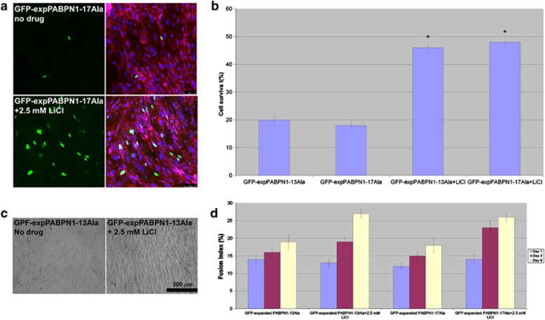
Expansion of polyalanine tracts causes at least nine inherited human diseases. Among these, a polyalanine tract expansion in the poly (A)-binding protein nuclear 1 (expPABPN1) causes oculopharyngeal muscular dystrophy (OPMD). So far, there is no treatment for OPMD patients. Developing drugs that efficiently sustain muscle protection by activating key cell survival mechanisms is a major challenge in OPMD research. Proteins that belong to the Wnt family are known for their role in both human development and adult tissue homeostasis. A hallmark of the Wnt signaling pathway is the increased expression of its central effector, beta-catenin (β-catenin) by inhibiting one of its upstream effector, glycogen synthase kinase (GSK)3β. Here, we explored a pharmacological manipulation of a Wnt signaling pathway using lithium chloride (LiCl), a GSK-3β inhibitor, and observed the enhanced expression of β-catenin protein as well as the decreased cell death normally observed in an OPMD cell model of murine myoblast (C2C12) expressing the expanded and pathogenic form of the expPABPN1. Furthermore, this effect was also observed in primary cultures of mouse myoblasts expressing expPABPN1. A similar effect on β-catenin was also observed when lymphoblastoid cells lines (LCLs) derived from OPMD patients were treated with LiCl. We believe manipulation of the Wnt/β-catenin signaling pathway may represent an effective route for the development of future therapy for patients with OPMD.


Figures

Figure 1
Figure 1
Choosing the best concentration of LiCl on the C2C12 cell line. LiCl (GSK inhibitor) at 2.5 mM maintains growth, size, and proliferation of C2C12 muscle cells. A dose-response experiment on non-transfected C2C12 cells shows that with a low dose of 2.5 mM LiCl, the cells maintain their growth and survival similarly to untreated cells. Cells were monitored under a live-stage Leica DMI 6000 microscope for morphology and viability. Phase images were captured at the indicated days after LiCl treatment. Higher concentrations of LiCl at 5 and 15 mM (top two rows) were toxic and caused the cells to die, while a concentration of 2.5 mM LiCl maintained the cells healthy and in good shape for 6 days. Therefore, 2.5 mM LiCl was the chosen concentration throughout the study
Figure 2
Figure 2
LiCl rescues GFP-expPABPN (13Ala and 17Ala)-associated cell death. (a) Cell survival determined by live-stage microscopy. C2C12 cells were transfected with a GFP vector, GFP-wtPABPN1-10Ala, GFP-expPABPN1-13Ala, and GFP-expPABPN1-17Ala, treated or not with 2.5 mM LiCl. The cells were counted every 24 h post transfection for 6 days consecutively using the live-stage microscope. The percentage of transfected living cells represents the variation of the amount of transfected living cells at different time points compared with the number of transfected cells obtained on day 1. LiCl rescues GFP-expPABPN1-13Ala and 17Ala-associated cell death (*P<0.001 versus non-treated samples). Mean±S.E., *P<0.05 compared with any other groups (ANOVA analysis). The experiment was repeated 3 times. (b) Cell death measured by fluorescent flow cytometry (FFC). Percentage of cell death observed by two-color FFC analysis with 7AAD on day 6 post-treatment with 2.5 mM LiCl. GFP-wtPABPN1-10Ala and GFP were used as controls. Cell death was calculated by dividing the number of 7AAD-stained transfected C2C12 cells over the total number of transfected cells. The experiment was repeated four times. (*P<0.001 versus non-treated samples). (c) A two-color FACS dotplot from a representative experiment in which C2C12 cells were transfected with GFP-expPABPN1-17Ala and treated with 2.5 mM LiCl (bottom) compared with non-treated cells (top), stained with 7AAD to label dead cells, and co-sorted for GFP (green) and 7AAD (red) fluorescence. Grid lines were positioned after calibrating the flow cytometry. Upper right quadrants signify 7AAD-labeled dead or dying transfected cells (GFP+7AAD+), i.e., Q2 (underlined values)
Figure 3
Figure 3
LiCl enhances cell proliferating and differentiation of C2C12 cells transfected with GFP-expPABPN1-17Ala. (a and b) The effect of LiCl treatment on OPMD C2C12 cell model controls. LiCl maintains the ability of cells expressing GFP-wtPABPN1-10Ala (a) and GFP (b) to proliferate and differentiate over the time course. Cells were transiently transfected with GFP-wtPABPN1-10Ala or GFP and treated with 2.5 mM LiCl for 6 days in DM. (c) LiCl protects against cell death and enhances the number of multinucleated differentiated myotubes as well as increasing their diameter's size (bottom panels). Cells were transiently transfected with GFP-expPABPN1-17Ala and treated with 2.5 mM LiCl for 6 days in DM. Cells were visualized daily under the live-stage microscope for morphology and viability. Representative images were captured on day 2 and day 6 post-treatment with the drug. (d) LiCl enhances muscle differentiation of C2C12 cells expressing GFP-expPABPN1-13Ala. Representative images showing the effect of LiCl on cell differentiation. LiCl increases the total number of multinucleated myotubes in cells expressing GFP-expPABPN1-13Ala (bottom left) compared with non-treated GFP-expPABPN1-13Ala cells (top left). Phase contrast image of myotubes treated with 2.5 mM LiCl (bottom right) shows the increased diameter size when compared with non-treated cells (top right) on day 6 after differentiation. White arrows point towards the myotube's diameter. Red arrowheads point towards dead cells or debris of dead cells
Figure 4
Figure 4
LiCl enhances muscle differentiation of C2C12 cells expressing GFP-expPABPN1-13Ala and 17Ala. LiCl increases the differentiation index of C2C12 cells expressing GFP-expPABPN1 (13Ala and 17Ala). The histogram represents the fusion index calculated at the indicated times after the addition of DM for cells expressing GFP-expPABPN1-13Ala and GFP-expPABPN1-17Ala treated or not treated with 2.5 mM LiCl. Myogenic index was determined as the ratio of the nuclei number of cells containing two or more nuclei to the total nuclei number of desmin C2C12-stained myotubes. The number of nuclei within each individual myotube was counted for 100–150 myotubes per well. At least three different fields containing 100 cells were counted. Data are the mean ±S.E. of three independent experiments
Figure 5
Figure 5
LiCl increases cell survival and differentiation of primary cultures of mouse myoblasts expressing GFP-expPABPN1 (13Ala and 17Ala). (a) Cell survival determined by live-stage microscopy. Confocal images showing the presence of an increased number of green cells (GFP-expPABPN1-17Ala) following a treatment (2.5 mM LiCl, bottom) support their increased survival by comparison with what is observed when the same cells were not treated with LiCl (top). Immnocytochemistry images were taken at day 6 post-transfection. GFP-expPABPN1-17Ala (green), desmin (red), and DAPI (blue). (b) LiCl increases the number of viable cells. Primary cultures mouse myoblasts expressing GFP-expPABPN1-13Ala, and GF-expPABPN1-17Ala, treated or not with 2.5 mM LiCl were counted at day 6 post-transfection using the live-stage microscope. Three different fields were chosen and cells were counted. Mean±S.E., *P<0.05 compared with any other groups (ANOVA analysis). The experiment was repeated three times. (c) LiCl enhances muscle differentiation of primary mouse myoblasts expressing GFP-expPABPN1-13Ala. Representative images showing the effect of LiCl on cell differentiation. Phase contrast images of myotubes treated with 2.5 mM LiCl (right) show an increased number of many elongated myotubes compared with non-treated cells (left) on day 6 after differentiation. (d) LiCl increases the differentiation index of primary mouse myoblast cells expressing GFP-expPABPN1 (13Ala and 17Ala). The histogram represents the fusion index calculated at the indicated times after DM addition for cells expressing GFP-expPABPN1-13Ala and GFP-expPABPN1-17Ala treated or not with 2.5 mM LiCl. At least three different fields containing 100 cells were counted. Data are the mean±S.E. of three independent experiments
Figure 6
Figure 6
LiCl leads to a significant increase of the β-catenin level in the OPMD C2C12 cell model. (a and b) LiCl leads to a significant increase of β-catenin in the OPMD C2C12 cell model while leaving the GFP level not affected. A western blot showing the effect of LiCl treatment on the β-catenin level at 48 h post transfection. C2C12 myoblast cells were transiently transfected with different PABPN1constructs as well as GFP, and non-transfected cells, and treated or not with two different doses of LiCl as indicated. Actin antibody was used to confirm equal loading. (c and d) LiCl treatment results in re-distribution of β-catenin from a cytoplasmic to a nuclear compartment. Confocal images of immunocytochemistry showing the subcellular distribution of β-catenin. A clear accumulation of β-catenin in the nucleus was observed in the C2C12-transfected cells with GFP-wtPABPN1-10Ala (c) and GFP-expPABPN1-17Ala (d) after LiCl treatment. GFP-PABPN1 constructs (green), β-catenin (red)
Figure 7
Figure 7
Increased level of β-catenin protein in the OPMD patient LCLs upon LiCl treatment. (a and b) show no difference in the β-catenin level between OPMD and control LCLs. (a) A Western blot of proteins extracted from controls and OPMD LCLs and detected with the β-catenin antibody. Actin is used as a control for equal loading of protein. The two control LCLs are: 39 196 and 34 299. The two patient OPMD LCLs are: 34 265 and 34 263. (b) Confocal images showing similar distribution of β-catenin protein in both control and patient OPMD LCLs. Immunofluorescent images of LCLs from control 34 299 (top panels) and patient 34 262 (bottom panel) were detected using the β-catenin antibody (green). TOTO (blue) stains for the nucleus. LCLs from control and OPMD patient are 34 299 and 34 262, respectively. (c) The western blot shows increased level of β-catenin protein after 48 h of LiCl treatment. The numbers refer to different OPMD LCLs. Actin was used to confirm equal loading of protein

References

    1. Dion P, Shanmugam V, Gaspar C, Messaed C, Meijer I, Toulouse A, et al. Transgenic expression of an expanded (GCG)13 repeat PABPN1 leads to weakness and coordination defects in mice. Neurobiol Dis. 2005;18:528–536. - PubMed
    1. Boukriche Y, Maisonobe T, Masson C. Neurogenic involvement in a case of oculopharyngeal muscular dystrophy. Muscle Nerve. 2002;25:98–101. - PubMed
    1. Schober R, Kress W, Grahmann F, Kellermann S, Baum P, Gunzel S, et al. Unusual triplet expansion associated with neurogenic changes in a family with oculopharyngeal muscular dystrophy. Neuropathology. 2001;21:45–52. - PubMed
    1. Brais B, Rouleau GA, Bouchard JP, Fardeau M, Tome FM. Oculopharyngeal muscular dystrophy. Semin Neurol. 1999;19:59–66. - PubMed
    1. Brais B, Bouchard JP, Xie YG, Rochefort DL, Chretien N, Tome FM, et al. Short GCG expansions in the PABP2 gene cause oculopharyngeal muscular dystrophy. Nat Genet. 1998;18:164–167. - PubMed

Publication types

MeSH terms