. 2013 Oct 3;4(10):e828.
doi: 10.1038/cddis.2013.337.

Cancer-initiating cells derived from human rectal adenocarcinoma tissues carry mesenchymal phenotypes and resist drug therapies

Affiliations

Cancer-initiating cells derived from human rectal adenocarcinoma tissues carry mesenchymal phenotypes and resist drug therapies

C-W Fan et al. Cell Death Dis. .

Abstract

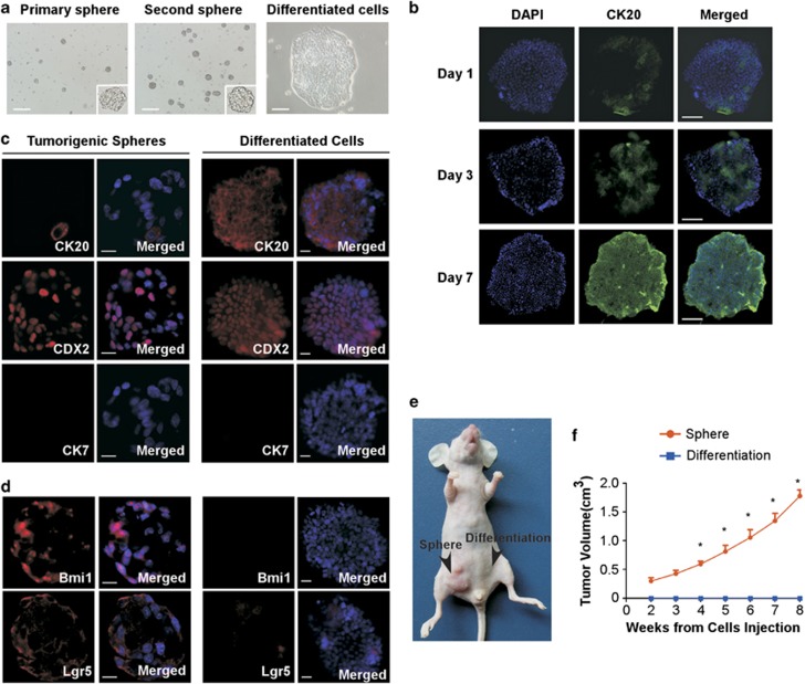
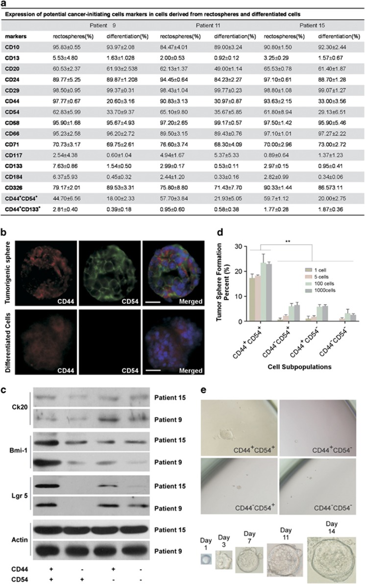
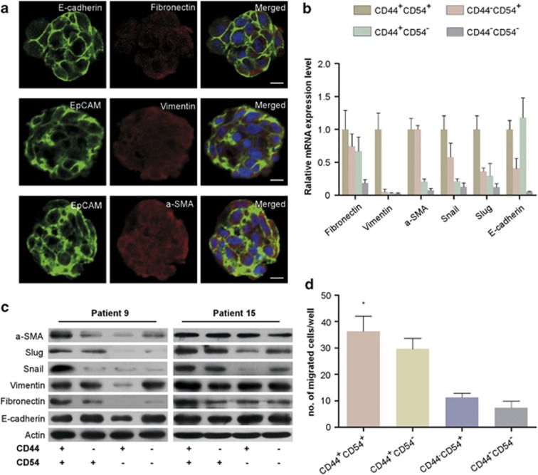
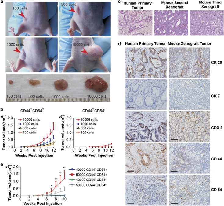
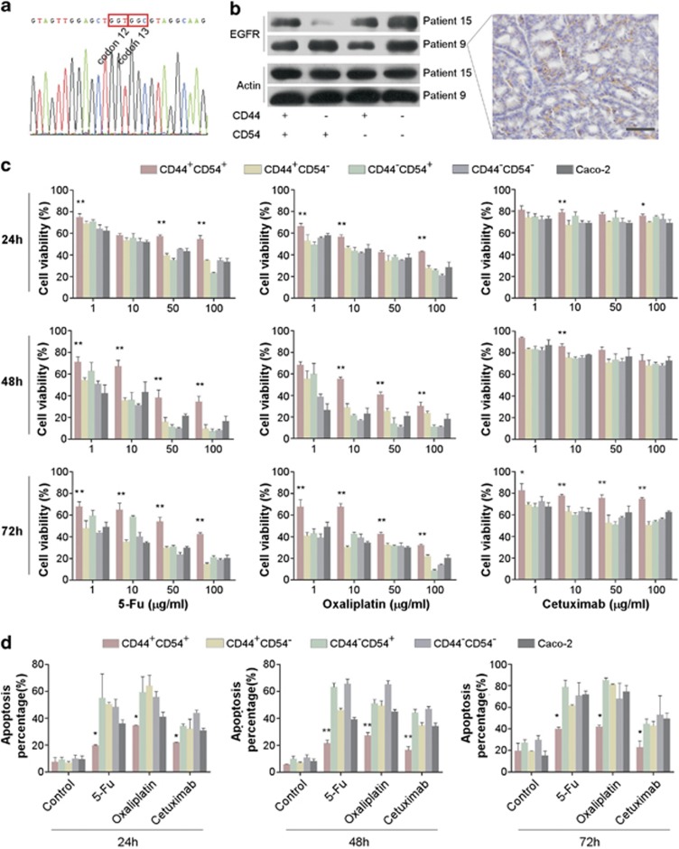
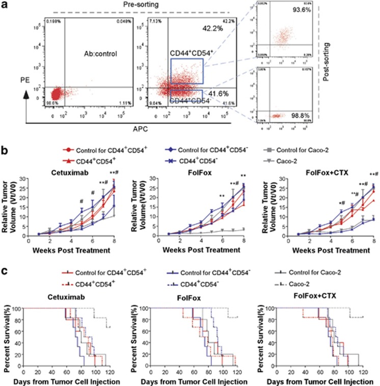
Accumulating evidence indicates that cancer-initiating cells (CICs) are responsible for cancer initiation, relapse, and metastasis. Colorectal carcinoma (CRC) is typically classified into proximal colon, distal colon, and rectal cancer. The gradual changes in CRC molecular features within the bowel may have considerable implications in colon and rectal CICs. Unfortunately, limited information is available on CICs derived from rectal cancer, although colon CICs have been described. Here we identified rectal CICs (R-CICs) that possess differentiation potential in tumors derived from patients with rectal adenocarcinoma. The R-CICs carried both CD44 and CD54 surface markers, while R-CICs and their immediate progenies carried potential epithelial-mesenchymal transition characteristics. These R-CICs generated tumors similar to their tumor of origin when injected into immunodeficient mice, differentiated into rectal epithelial cells in vitro, and were capable of self-renewal both in vitro and in vivo. More importantly, subpopulations of R-CICs resisted both 5-fluorouracil/calcium folinate/oxaliplatin (FolFox) and cetuximab treatment, which are the most common therapeutic regimens used for patients with advanced or metastatic rectal cancer. Thus, the identification, expansion, and properties of R-CICs provide an ideal cellular model to further investigate tumor progression and determine therapeutic resistance in these patients.


Figures

Figure 1
Figure 1
The differentiation potential and tumorigenic capacity of rectospheres in vitro. (a) Example of tumor spheres generated from a human rectal cancer sample that were passaged twice and differentiated cell. Primary spheres: spheres directly generated from human tumor tissues. Secondary spheres: spheres representing the second passage of primary spheres. Bars=500 μM. Differentiated cells: spheroids were induced by 20% fetal bovine serum. One representative of four independent spheroid cultures is shown. Bars=50 μM. (b) Spheres were cultured in SCM and analyzed by immunofluorescence to detect the differentiation marker CK20 at different time points. Bars=50 μM. (c) Immunofluorescent analysis using CK20 (red), CDX2 (red), CK7 (red), and DAPI (4,6-diamidino-2-phenylindole; blue) stains. One representative image of three different tumors is shown. (d) Bmi1 and Lgr5 expressed on cells from rectospheres (left panels) and rectosphere-derived differentiation progeny (right panels). Nuclei were counterstained by DAPI (blue). One representative experiment of three different tumors is shown. Bars=25 μM. (e) The example shows that spheres cultured in SFM, but not in SCM, recapitulate tumors in nude mice. (f) Tumorigenic potential of tumor spheres after subcutaneous injection. Size of subcutaneous rectal carcinoma tumors generated from 103 sphere cells and 1 × 10⁶ differentiated cells. Data are mean±S.D. of two independent experiments, each performed with cells from different donors (*P<0.01; patients 9 and 15)
Figure 2
Figure 2
CD44+CD54+cells derived from rectospheres possess strong self-renewal capability. (a) Flow cytometry analysis of putative CIC markers on rectospheres and rectosphere-derived differentiation progeny in three independent experiments (patients 9, 11, and 15). (b) Immunofluorescence analysis using antibodies against CD44 (red) and CD54 (green) on cells from rectal cancer spheres (upper panels) and differentiated rectal cancer cells (lower panels). Nuclei were counterstained by DAPI (4,6-diamidino-2-phenylindole; blue). One representative image of two different tumors is shown. Bars=50 μm. (c) Immunoblotting validation of stemness and differentiation gene expression in different cellular populations derived from two independent donors (patients 9 and 15). (d) Tumor sphere formation ratio derived from different number of CD44+CD54+, CD44+CD54, CD44CD54+, and CD44CD54 cells in SFM. Data are mean±S.D. of three independent experiments, each performed with cells from different donors (**P<0.01; patients 9, 11 and 15). (e) Single CD44+CD54+ cell generated one sphere in culture (left upper panel). Phase-contrast microscopy representative images taken at different time points confirmed spheroid growth from single-sorted CD44+CD54+ cells. One representative experiment of two different tumors is shown
Figure 3
Figure 3
Rectal cancer sphere cells exhibit mesenchymal phenotypes. (a) Confocal images of rectospheres stained with antibodies against E-cadherin (green), EpCAM (green), fibronectin (red), vimentin (red), and α-SMA (α-smooth muscle actin; red). One representative image of two different tumors is shown. Bar=25 μM. (b) Expression levels of mRNAs encoding E-cadherin, Snail, Slug, fibronectin, vimentin, and α-SMA in different cellular subpopulations were determined by real-time reverse transcriptase-PCR. Data are mean±S.D. of three independent experiments, each performed with cells from different donors (patients 9, 11, and 15). (c) Immunoblotting of EMT-related proteins from lysates of different cell populations isolated from rectospheres of two different patients (patients 9 and 15). (d) Migration capacity of different cellular subpopulations. Data are mean±S.D. of three independent experiments, each performed with cells from different donors (*P<0.05; patients 9, 11, and 15)
Figure 4
Figure 4
CD44+CD54+ cells derived from rectospheres have the strongest tumorigenicity among the four cellular subpopulations. (a) Tumor-bearing mice derived from 100, 500, 1000, and 10 000 CD44+CD54+ rectal cancer cells and excised subcutaneous tumors. One representative experiment of three different tumors is shown. (b) Size of xenografts of CD44+CD54+ and CD44+CD54 derived from rectospheres. Data are mean tumor size±S.D. of 3–5 tumors per group derived from three separate patients (patients 9, 11, and 15). (c) Hematoxylin and eosin analysis of a human rectal cancer section from the original human tumor and corresponding xenografts obtained after injection of sphere cells. Bars=100 μM. Second and third xenografts: tumors were obtained from the first and second transplantation, respectively. One representative experiment of two different tumors is shown. (d) Immunohistochemical analysis of relative differentiation and putative R-CIC markers in tissue derived from original human tumor (left panel) and sphere-derived first xenografts (right panel). One representative experiment of two different tumors is shown. Bars=100 μM. (e) Size of xenografts of CD44+CD54+ and CD44+CD54 sorted from three rectal cancer tissues. Data are mean tumor size±S.D. of 3–5 tumors per group derived from three separate patients (**P<0.01, *P<0.05; patients 31, 32, and 33)
Figure 5
Figure 5
CD44+CD54+ R-CICs are resistant to chemotherapeutic agents and apoptosis in vitro. (a) Examination of k-ras gene mutation with sequencing, where codons 12 and 13 of exon 2 are wild type. One representative graph of three different tumors is shown. (b) Immunoblotting or immunohistochemical validation of EGFR expression in different cell populations derived from two independent experiments (patients 9 and 15). (c) Percentage of cell viability of different cellular subpopulations and caco-2 cells after treatment with 5-Fu, oxaliplatin, and cetuximab of different concentrations for 24, 48, and 72 h in vitro. Data are mean±S.D. of three independent experiments, each performed with cells from different donors (**P<0.01, *P<0.05; patients 9, 11, and 15). (d) Cell apoptosis percentage of different cellular subpopulations and caco-2 cells after treatment with 5-Fu, oxaliplatin, or cetuximab for 24, 48, or 72 h. Data are mean±S.D. of three independent experiments, each performed with cells from different donors (**P<0.01, *P<0.05; patients 9,11, and 15)
Figure 6
Figure 6
CD44+CD54+ R-CICs resist FolFox and cetuximab therapy in vivo. (a) The definition of CD44+CD54+ and CD44+CD54 cellular populations in rectospheroids and the resultant purity after cell sorting. (b) Treatment of tumor-bearing mice generated from CD44+CD54+ R-CICs, CD44+CD54, and caco-2 cells with cetuximab, FolFox, and cetuximab plus FolFox in vivo. Data are mean tumor size±S.D. of five tumors per group derived from two independent experiments from different patients. (#P<0.01, CD44+CD54 compared with its control; **P<0.01, *P<0.05; Caco2 cells compared with its control). (c) Survival of tumor-bearing mice derived from CD44+CD54+ R-CICs (P=0.684, cetuximab; P=0.824, FolFox; P=0.207, FolFox–cetuximab), CD44+CD54 (P=0.019, cetuximab; P=0.001, FolFox; P=0.067, FolFox–cetuximab), and caco-2 cells (P=0.003, cetuximab; P=0.003, FolFox; P=0.003, FolFox–cetuximab) treated with different regimens

References

    1. Jemal A, Siegel R, Xu J, Ward E. Cancer statistics. CA Cancer J Clin. 2010;2010;60:277–300. - PubMed
    1. Yamauchi M, Morikawa T, Kuchiba A, Imamura Y, Qian ZR, Nishihara R, et al. Assessment of colorectal cancer molecular features along bowel subsites challenges the conception of distinct dichotomy of proximal versus distal colorectum. Gut. 2012;61:847–854. - PMC - PubMed
    1. Yu H, Harris RE, Gao YT, Gao R, Wynder EL. Comparative epidemiology of cancers of the colon, rectum, prostate and breast in Shanghai, China versus the United States. Int J Epidemiol. 1991;20:76–81. - PubMed
    1. Cunningham D, Atkin W, Lenz HJ, Lynch HT, Minsky B, Nordlinger B, et al. Colorectal cancer. Lancet. 2010;375:1030–1047. - PubMed
    1. Ricci-Vitiani L, Fabrizi E, Palio E, De Maria R. Colon cancer stem cells. J Mol Med. 2009;87:1097–1104. - PubMed

Publication types

MeSH terms

Supplementary concepts