. 2013 Oct 3;4(10):e831.
doi: 10.1038/cddis.2013.347.

Epithelial-mesenchymal transition markers expressed in circulating tumor cells in hepatocellular carcinoma patients with different stages of disease

Affiliations

Epithelial-mesenchymal transition markers expressed in circulating tumor cells in hepatocellular carcinoma patients with different stages of disease

Y-M Li et al. Cell Death Dis. .

Abstract

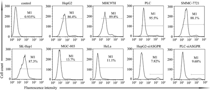
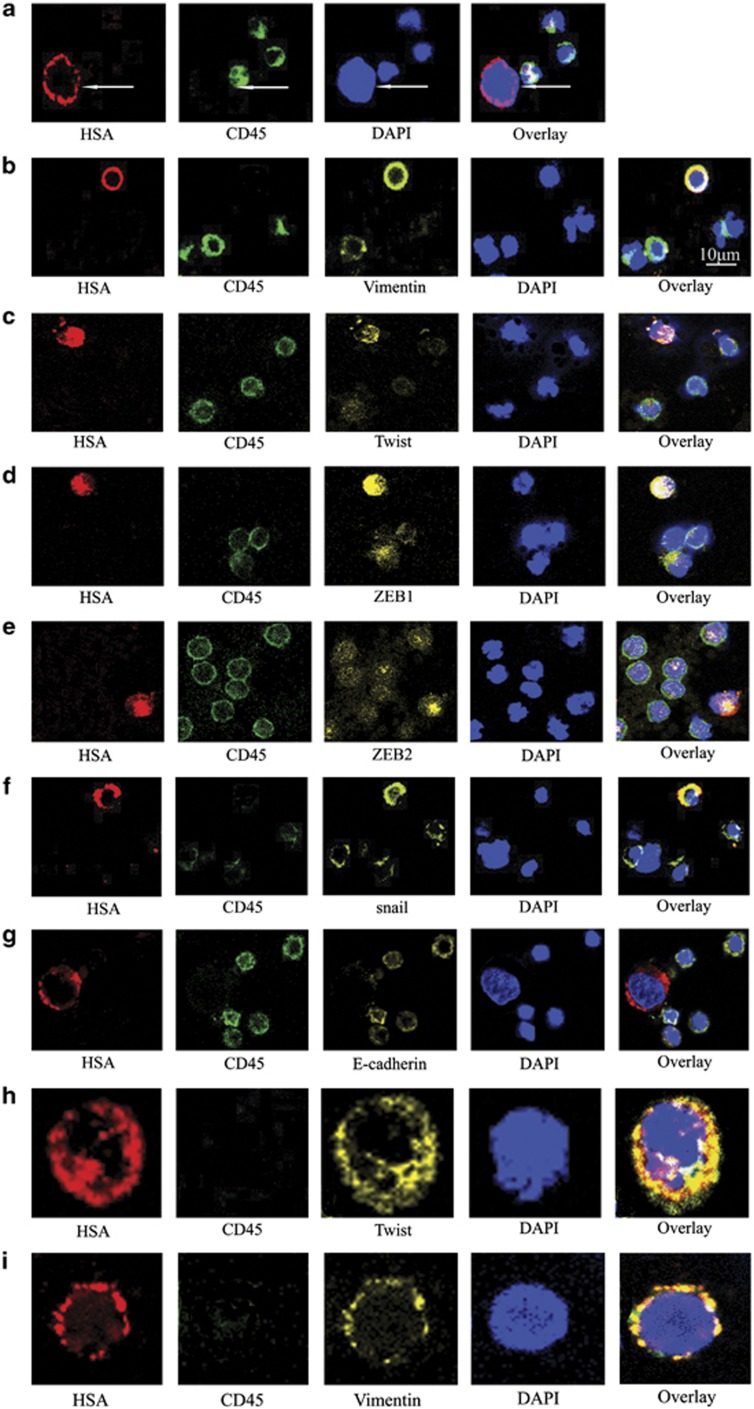
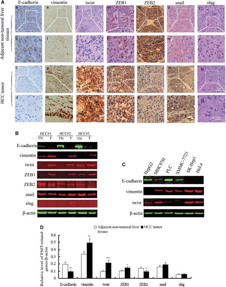
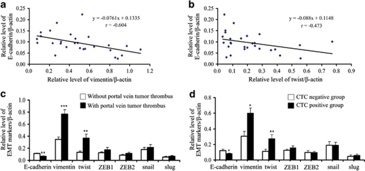
The presence of circulating tumor cells (CTCs) in peripheral blood is associated with metastasis and prognosis in hepatocellular carcinoma (HCC) patients. The epithelial-mesenchymal transition (EMT) has a pivotal role in tumor invasion and dissemination. To identify more sensitive biomarkers for evaluating metastasis and prognosis, we investigated the expression of EMT markers, including vimentin, twist, ZEB1, ZEB2, snail, slug and E-cadherin in CTCs, primary HCC tumors and adjacent non-tumoral liver tissues. After isolating viable CTCs from the peripheral blood of HCC patients using asialoglycoprotein receptors (ASGPRs), the CTCs were identified with immunofluorescence staining. CTCs were detected in the peripheral blood obtained from 46 of 60 (76.7%) HCC patients. Triple-immunofluorescence staining showed that twist and vimentin expression could be detected in CTCs obtained from 39 (84.8%) and 37 (80.4%) of the 46 patients, respectively. The expression of both twist and vimentin in CTCs was significantly correlated with portal vein tumor thrombus. Coexpression of twist and vimentin in CTCs could be detected in 32 (69.6%) of the 46 patients and was highly correlated with portal vein tumor thrombus, TNM classification and tumor size. Quantitative fluorescence western blot analysis revealed that the expression levels of E-cadherin, vimentin and twist in HCC tumors were significantly associated with the positivity of isolated CTCs (P=0.013, P=0.012, P=0.009, respectively). However, there was no significant difference in ZEB1, ZEB2, snail and slug expression levels in CTCs, primary HCC tumors and adjacent non-tumoral liver tissues across samples with regard to the clinicopathological parameters. Our results demonstrate that the EMT has a role in promoting the blood-borne dissemination of primary HCC cells, and the twist and vimentin expression levels in CTCs could serve as promising biomarkers for evaluating metastasis and prognosis in HCC patients.


Figures

Figure 1
Figure 1
Flow cytometric analysis confirmed the specific binding of biotinylated asialofetuin to ASGPR. HepG2, MHC97H, PLC, SMMC-7721 and SK-Hep1 are human hepatoma cell lines expressing ASGPR. The human gastric cancer cell line MGC-803 and the HeLa human cervical adenocarcinoma cell lines are both negative for ASGPR. All the cells were stained using biotinylated asialofetuin and antibiotin-luorescein isothiocyanate (FITC), excluding the HepG2 cells stained with PBS and antibiotin-FITC, which served as the controls. After ASGPR was knocked down in HepG2 and PLC cells, biotinylated asialofetuin binding was barely detectable in these two cell lines
Figure 2
Figure 2
Immunofluorescence staining identified the expression of EMT-related genes in CTCs obtained from HCC patients with different stages of disease. (a) CTCs and hematologic cells were stained with the HSA anti-mouse antibody/Alexa Fluor 647 rabbit anti-mouse IgG (red) and CD45 anti-rat antibody/Alexa Fluor 488 rabbit anti-rat IgG (green). The cell nuclei were stained with DAPI (blue). (bg) Triple-immunofluorescence shows CTCs expressing vimentin, twist, ZEB1, ZEB2, snail and E-cadherin, respectively. These CTCs were stained with primary antibodies raised in rabbit against vimentin, twist, ZEB1, ZEB2, snail and E-cadherin, and the corresponding secondary antibody was Alexa Fluor 555 donkey anti-rabbit IgG (yellow). The remaining staining procedures were as same as those described for double-immunofluorescence. Bar=10 μm. Twist and vimentin coexpression in CTCs obtained from the same patient. Representative photomicrographs of CTCs obtained from the same patient in different visual fields. (h) A CTC expressing HSA and twist but not CD45. (i) A CTC expressing HSA and vimentin but not CD45. Original magnification × 800
Figure 3
Figure 3
EMT-related markers are significantly differentially expressed in primary HCC tumors compared with adjacent non-tumoral tissues. (A) E-cadherin was highly expressed in adjacent non-tumoral liver tissues (a, b), and the expression of E-cadherin was nearly absent in tumor tissues (c, d). Vimentin, twist and ZEB1 expression was significantly lower in adjacent non-tumoral liver tissues (e, f; i, j; m, n) than in primary HCC tumors (g, h; k, l; o, p). The expression of ZEB2 was prone to be downregulated in HCC tumors (s, t) compared with adjacent non-tumoral liver tissues (q, r). There was no apparent difference in the snail expression in adjacent non-tumoral liver tissues (u, v) and HCC tumors (w, x). Slug expression was barely detectable in adjacent non-tumoral liver tissues (y, z) or HCC tumors (α, β). Original magnification × 100 (a, c, e, g, i, k, m, o, q, s, u, w, y, α), bar=100 μm. and corresponding areas with higher magnification × 200 (b, d, f, h, j, l, n, p, r, t, v, x, z, β), bar=50 μm. (B) A representative immunoblot of E-cadherin, vimentin, twist, ZEB1, ZEB2, snail and slug protein levels in adjacent non-tumoral liver tissues (TN) and HCC tumors (T). β-actin was used as an internal standard for protein loading. (C) A representative immunoblot of E-cadherin, vimentin and twist expression in several HCC cell lines and HeLa cells. (D) The relative quantification analysis revealed that E-cadherin was significantly downregulated in HCC tumors compared with the adjacent non-tumoral liver tissues (n=28, **P=0.003). Vimentin, twist and ZEB1 expression levels were significantly upregulated in HCC tumors compared with the adjacent non-tumoral liver tissues (n=28, **P=0.002, ***P<0.001, *P=0.016). ZEB2 expression was significantly decreased in HCC tumors compared with the adjacent non-tumoral liver tissues (n=28, *P=0.030). There was no statistically significant difference for snail and slug expression in HCC tumors or paired adjacent non-tumoral liver tissues
Figure 4
Figure 4
The occurrence of the EMT in HCC correlated with portal vein tumor thrombus and promoted CTC formation. (a) The relative level of vimentin expression was negatively correlated with E-cadherin expression in HCC tumors (n=28, r=−0.604, P=0.001). (b) The relative level of twist expression was negatively correlated with E-cadherin expression in HCC tumors (n=28, r=−0.437, P=0.020). (c) The expression of E-cadherin in HCC tumors was significantly lower in patients with portal vein tumor thrombus (n=10) than in patients without portal vein tumor thrombus (n=18) (**P=0.007), vimentin and twist expression in HCC tumors was significantly higher in patients with portal vein tumor thrombus (n=10) than in patients without portal vein tumor thrombus (n=18) (***P<0.001, **P=0.004, respectively). There was no significant correlation between the expression of other transcriptional factors and portal vein tumor thrombus. (d) The expression of E-cadherin in HCC tumors was significantly lower in the CTC-positive patients (n=18) than in the CTC-negative patients (n=10) (*P=0.013). The expression of vimentin and twist in HCC tumor was significantly higher in CTC-positive patients (n=18) than in the CTC-negative patients (n=10) (*P=0.012, **P=0.009, respectively). The expression levels of the other transcriptional factors were not correlated with the positivity rate of CTCs

References

    1. El-Serag HB, Rudolph KL. Hepatocellular carcinoma: epidemiology and molecular carcinogenesis. Gastroenterology. 2007;132:2557–2576. - PubMed
    1. Altekruse SF, McGlynn KA, Reichman ME. Hepatocellular carcinoma incidence, mortality, and survival trends in the United States from 1975 to 2005. J Clin Oncol. 2009;27:1485–1491. - PMC - PubMed
    1. Yoo HY, Patt CH, Geschwind JF, Thuluvath PJ. The outcome of liver transplantation in patients with hepatocellular carcinoma in the United States between 1988 and 2001: 5-year survival has improved significantly with time. J Clin Oncol. 2003;21:4329–4335. - PubMed
    1. Ding W, You H, Dang H, LeBlanc F, Galicia V, Lu SC, et al. Epithelial-to-mesenchymal transition of murine liver tumor cells promotes invasion. Hepatology. 2010;52:945–953. - PMC - PubMed
    1. Vogel I, Kalthoff H. Disseminated tumour cells. Their detection and significance for prognosis of gastrointestinal and pancreatic carcinomas. Virchows Arch. 2001;439:109–117. - PubMed

Publication types

MeSH terms