. 2013 Dec 1;12(23):3615-28.
doi: 10.4161/cc.26588. Epub 2013 Oct 1.

Phosphorylation of eIF4GII and 4E-BP1 in response to nocodazole treatment: a reappraisal of translation initiation during mitosis

Affiliations

Phosphorylation of eIF4GII and 4E-BP1 in response to nocodazole treatment: a reappraisal of translation initiation during mitosis

Mark J Coldwell et al. Cell Cycle. .

Abstract

Translation mechanisms at different stages of the cell cycle have been studied for many years, resulting in the dogma that translation rates are slowed during mitosis, with cap-independent translation mechanisms favored to give expression of key regulatory proteins. However, such cell culture studies involve synchronization using harsh methods, which may in themselves stress cells and affect protein synthesis rates. One such commonly used chemical is the microtubule de-polymerization agent, nocodazole, which arrests cells in mitosis and has been used to demonstrate that translation rates are strongly reduced (down to 30% of that of asynchronous cells). Using synchronized HeLa cells released from a double thymidine block (G 1/S boundary) or the Cdk1 inhibitor, RO3306 (G 2/M boundary), we have systematically re-addressed this dogma. Using FACS analysis and pulse labeling of proteins with labeled methionine, we now show that translation rates do not slow as cells enter mitosis. This study is complemented by studies employing confocal microscopy, which show enrichment of translation initiation factors at the microtubule organizing centers, mitotic spindle, and midbody structure during the final steps of cytokinesis, suggesting that translation is maintained during mitosis. Furthermore, we show that inhibition of translation in response to extended times of exposure to nocodazole reflects increased eIF2α phosphorylation, disaggregation of polysomes, and hyperphosphorylation of selected initiation factors, including novel Cdk1-dependent N-terminal phosphorylation of eIF4GII. Our work suggests that effects on translation in nocodazole-arrested cells might be related to those of the treatment used to synchronize cells rather than cell cycle status.

Keywords: 4E-BP1; Cdk1; cell cycle; eIF4GII; eukaryotic translation initiation factor; nocodazole.


Figures

None
Figure 1. Protein synthesis rates are not decreased during M phase when observing cycling synchronized cells. (A) Exponentially growing HeLa cells were maintained as an asynchronous population or synchronized at the G1/S boundary by double thymidine block and released for the number of hours indicated. Nuclei content of cells at each time point was determined by staining with propidium iodide and FACS analysis. (B) In parallel experiments, translation rates were examined by pulsing cells with [35S] methionine for 30 min prior to harvest. Extracts were prepared, and the incorporation of radioactive methionine into protein (cpm/μg total protein) determined as described in the text. For the T/N sample, HeLa cells were blocked once in thymidine before being released then synchronized in M phase by incubation with the microtubule depolymerizing agent nocodazole for 19 h. (C) Equal amounts of extract from (B) were subjected to SDS-PAGE and proteins transferred to PVDF. The membrane was then probed with the antibodies shown and visualized with secondary antibodies conjugated to HRP.
None
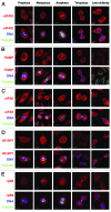
Figure 2. The translation machinery co-stains with the microtubule network during different stages of mitosis and cytokinesis. Cells released from a double thymidine block for 9 h were fixed with 4% (w/v) paraformaldehyde and permeabilized with 0.1% (v/v) Triton X-100. Cells in different stages of mitosis and cytokinesis were examined by confocal microscopy, as determined by the positioning of microtubules or DNA (A–E). Upper panels show components of the translation machinery which were detected with rabbit polyclonal antibodies, as indicated, followed by an anti-rabbit secondary antibody conjugated to Alexa Fluor 555 (red). Lower panels show this signal merged with α-tubulin detected with a mouse monoclonal antibody conjugated to FITC (green) and DNA detected with DAPI (blue).
None
Figure 3. Synchronization with nocodazole inhibits translation and leads to substantial phosphorylation of components of the translation initiation machinery. (A) HeLa cells were either maintained asynchronously, or blocked once in thymidine before being released, then synchronized in M phase by incubation with nocodazole for 19 h. As before, FACS analysis was used to determine nuclear DNA content. (B) Samples were subjected to sucrose density gradient centrifugation to determine the effect of incubation with nocodazole on polysome formation. Translation rates from these cells are shown as part of Figure 1 (B), which confirm the large inhibition of translation, as determined by the reduction in [35S] methionine incorporation. (C) Equal amounts of cell extracts from parallel samples were analyzed by immunoblotting for the proteins indicated. To separate isoforms of eIF4G proteins that arise from alternative translation initiation, low-percentage SDS-PAGE was used. (D) To determine whether shifts in migration of eIF4GI/II and 4E-BP1 were due to phosphorylation, extracts from asynchronous cells or cells which had been incubated with nocodazole for 19 h were prepared. These were then incubated in the presence or absence of lambda protein phosphatase for 30 min before the treated extracts were separated by SDS-PAGE and immunoblotted for the indicated proteins.
None
Figure 4. Robust phosphorylation of eIF2α does not occur following release from G2/M block, and arresting synchronous cells with nocodazole has negligible effects on translation rates. (A) HeLa cells were maintained asynchronously, subjected to a single thymidine nocodazole block, or incubated with the Cdk1 inhibitor RO3306 for 20 h at a concentration of 9 μM to arrest cells at the end of G2. As this latter inhibitor is reversible, the drug was washed off, and the cells progressed through mitosis, as observed by FACS analysis every 30 min. (B) Translation rates (obtained by pulsing for 15 min prior to harvest) were obtained from parallel experiments to (A). (C) Mobility shifts of eIF4GI and 4E-BP1 and the phosphorylation status of eIF2α were determined from cell extracts prepared in (B) by SDS-PAGE and immunoblotting with antibodies raised against standard or phospho-epitopes, as indicated. (D) Cells were synchronized with a double thymidine block as before, then released into medium which was either normal, supplemented with nocodazole at release, or supplemented with nocodazole 6 h after release. FACS analysis of cells at the time points indicated was used to show the arrest in G2/M in the cells incubated with nocodazole. (E) The amount of incorporation of [35S] methionine into protein for the 15 min prior to harvest was determined from parallel experiments to (D) as previously described. (F) As before, cell extracts from parallel experiments to (D) were made and subjected to SDS-PAGE and immunoblotting to show the phosphorylation status of eIF2α and 4E-BP1 in the 3 different treatments. (G) In an attempt to determine the novel phosphorylation site of 4E-BP1 that is detected following nocodazole treatment, the open reading frame of the human protein was inserted into a vector in frame with an N-terminal myc-tag and C-terminal 3× FLAG tag. Further variants were constructed where putative phosphorylation sites (S83 and S112) were mutated to alanine either singly or in combination. The 4 variants were then transfected into HeLa cells and either maintained asynchronously or treated with nocodazole for 19 h. Cell extracts were prepared and the migration of the exogenous proteins determined by SDS-PAGE, detected with anti-FLAG antibody. (H) As for (G) mutants of 4E-BP1 were prepared where putative phosphorylation sites (T82 and S83) were mutated to alanine. After transfection into HeLa cells and nocodazole treatment as before, migration of FLAG-tagged proteins was examined by immunoblotting.
None
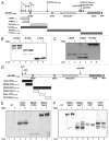
Figure 5. Deletion analysis of eIF4G proteins reveals two novel phosphoserine sites in eIF4GII. (A) A schematic representation of eIF4GI, showing the alternative AUG translation initiation sites (f through a) and the caspase-3 cleavage sites used to generate deletion fragments of this protein. Numbering conforms to the longest isoform as independently identified by others., Each cDNA was inserted into a vector containing an N-terminal myc-tag and C-terminal 3xFLAG tag to enable detection. (B) Plasmids containing cDNAs representing the 3 fragments of apoptotic cleavage of eIF4GI (FAGs) were transfected into HeLa cells either untreated or incubated with nocodazole for 19 h. Any changes in migration of the fragments due to the presence of nocodazole was determined by SDS-PAGE and immunoblotting using anti-FLAG antibody to detect fragment expression. (C) Further deletion fragments of N-FAGf were constructed as before to delineate regions likely to be phosphorylated, these were transfected into HeLa cells and migration in the presence or absence of nocodazole was examined. (D) Deletion fragments of the N-terminus of eIF4GII were constructed as before, in order to map the phosphorylation site(s) of this protein. The amino acid numbering conforms to the CUGb initiated isoform previously identified by our group, which extends the open reading frame N-terminally from the initially published AUG. (E) Migration of the N-terminal fragments was examined following treatment with nocodazole for 19 h, with proteins detected with anti-FLAG antibody. (F) Further subfragments were made of the eIF4GII open reading frame, as indicated in (D), and their migration in cells treated with nocodazole determined as before.
None
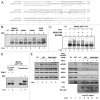
Figure 6. Identification of phosphorylation of eIF4GII at S385 and S389 by Cdk1, and consequences for initiation factor complex formation. (A) Mass spectroscopy was used to identify phosphorylation of two serine residues (384 and 392) within the MSGG-APPT fragment of eIF4GII following immunoprecipitation of the fragment from cells treated with nocodazole. The sites are shown in bold and underlined, and the surrounding sequences were aligned with eIF4GI using CLUSTALW, showing the poor conservation of this particular region of the protein. The site of PABP binding is shown in gray, with a dashed underline. The symbols *: and . respectively denote identical residues, or conserved and semi-conserved substitutions in the alignment. (B) Ser–Ala mutants, either single or in combination, of the MSGG-APPT fragment of eIF4GII were made and inserted into the myc-3× FLAG vector as before. Cells transfected with these vectors were incubated with or without nocodazole and migration of the proteins examined as before. (C) Prediction software (GPS 2.1.2) indicated that likely kinases responsible for this phosphorylation were p38 MAPK or Cdk1. Therefore cell extracts were made from cells transfected with MSGG-APPT that had been incubated with or without nocodazole and cell-permeable kinase inhibitors. These would either block p38 MAPK signaling (SB202190) or Cdk1 signaling (RO3306). Migration of the eIF4GII fragment was determined as previously described. (D) To confirm the results from panel (C), immunoprecipitated MSGG-AAPT protein was incubated in the presence of recombinant Cdk1/cyclin B and ATP. This was run alongside a positive control of extract from cells treated with or without nocodazole. (E) To determine whether the novel phosphorylation sites have any influence on the interactions of eIF4GII with other components of the translation initiation factor machinery, the S384/392A double mutant was introduced into the full-length eIF4GII CUGb ORF. This vector contains an N-terminal myc tag, and therefore exogenous eIF4GII could be immunoprecipitated from transfected cells which had been incubated with nocodazole. The left hand panel shows the levels of endogenous proteins as determined by SDS-PAGE and immunoblotting, and the right hand panel shows the eluate from a co-immunoprecipitation where myc-9E10 antibody had been captured on agarose resin.

References

    1. Jackson RJ, Hellen CU, Pestova TV. The mechanism of eukaryotic translation initiation and principles of its regulation. Nat Rev Mol Cell Biol. 2010;11:113–27. doi: 10.1038/nrm2838. - DOI - PMC - PubMed
    1. Laplante M, Sabatini DM. mTOR signaling in growth control and disease. Cell. 2012;149:274–93. doi: 10.1016/j.cell.2012.03.017. - DOI - PMC - PubMed
    1. Ma XM, Blenis J. Molecular mechanisms of mTOR-mediated translational control. Nat Rev Mol Cell Biol. 2009;10:307–18. doi: 10.1038/nrm2672. - DOI - PubMed
    1. Morley SJ, Coldwell MJ, Clemens MJ. Initiation factor modifications in the preapoptotic phase. Cell Death Differ. 2005;12:571–84. doi: 10.1038/sj.cdd.4401591. - DOI - PubMed
    1. Proud CG. mTOR Signalling in Health and Disease. Biochem Soc Trans. 2011;39:431–6. doi: 10.1042/BST0390431. - DOI - PubMed

Publication types

MeSH terms