

Molecular imaging in cardiovascular disease: Which methods, which diseases?
- PMID: 24092271
- PMCID: PMC3943209
- DOI: 10.1007/s12350-013-9785-0
Molecular imaging in cardiovascular disease: Which methods, which diseases?
Abstract
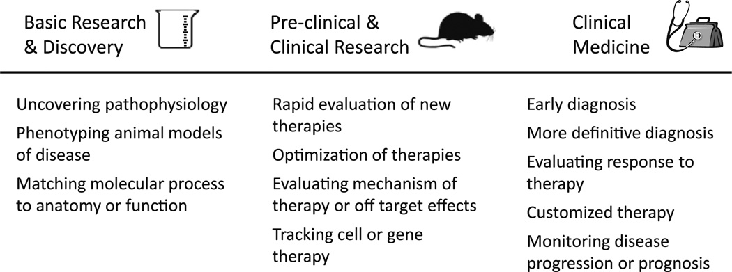
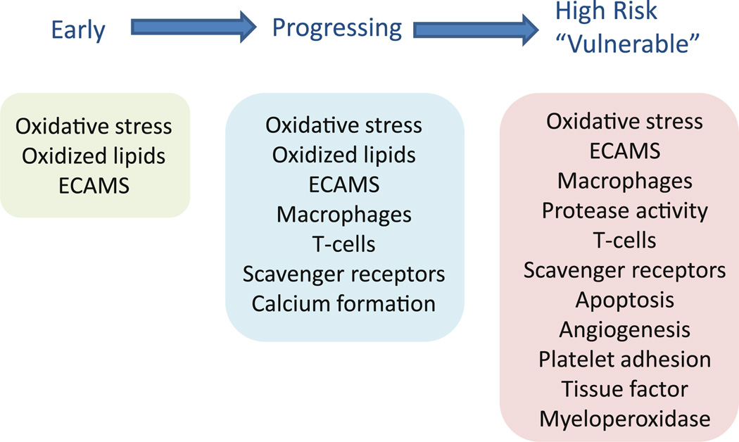
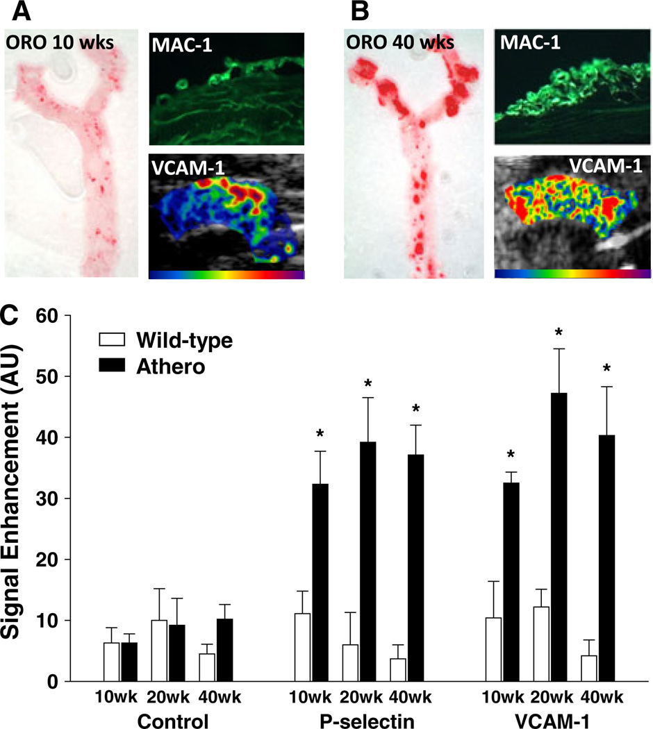
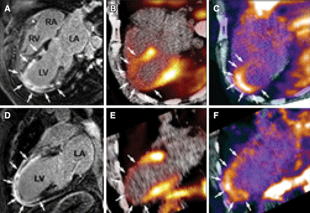
Techniques for in vivo assessment of disease-related molecular changes are being developed for all forms of non-invasive cardiovascular imaging. The ability to evaluate tissue molecular or cellular phenotype in patients has the potential to not only improve diagnostic capabilities but to enhance clinical care either by detecting disease at an earlier stage when it is more amenable to therapy, or by guiding most appropriate therapies. These new techniques also can be used in research programs in order to characterize pathophysiology and as a surrogate endpoint for therapeutic efficacy. The most common approach for molecular imaging involves the creation of novel-targeted contrast agents that are designed so that their kinetic properties are different in disease tissues. The main focus of this review is not to describe all the different molecular imaging approaches that have been developed, but rather to describe the status of the field and highlight some of the clinical and research applications that molecular imaging will likely provide meaningful benefit. Specific target areas include assessment of atherosclerotic disease, tissue ischemia, and ventricular and vascular remodeling.
Figures
References
-
- Kurt M, Shaikh KA, Peterson L, Kurrelmeyer KM, Shah G, Nagueh SF, et al. Impact of contrast echocardiography on evaluation of ventricular function and clinical management in a large prospective cohort. J Am Coll Cardiol. 2009;53:802–810. - PubMed
-
- Halpern EJ, Savage MP, Fischman DL, Levin DC. Cost-effectiveness of coronary CT angiography in evaluation of patients without symptoms who have positive stress test results. AJR Am J Roentgenol. 2010;194:1257–1262. - PubMed
-
- Ridker PM, Danielson E, Fonseca FA, Genest J, Gotto AM, Jr, Kastelein JJ, et al. Rosuvastatin to prevent vascular events in men and women with elevated C-reactive protein. N Engl J Med. 2008;359:2195–2207. - PubMed
-
- Yousuf O, Mohanty BD, Martin SS, Joshi PH, Blaha MJ, Nasir K, et al. High-sensitivity C-reactive protein and cardiovascular disease: A resolute belief or an elusive link? J Am Coll Cardiol. 2013;62:397–408. - PubMed
Publication types
MeSH terms
Grants and funding
LinkOut - more resources
Full Text Sources
Other Literature Sources
