

Peripancreatic cystic lymphangioma diagnosed by endoscopic ultrasound/fine-needle aspiration: a rare mesenchymal tumour
- PMID: 24092605
- PMCID: PMC3822152
- DOI: 10.1136/bcr-2013-200210
Peripancreatic cystic lymphangioma diagnosed by endoscopic ultrasound/fine-needle aspiration: a rare mesenchymal tumour
Abstract
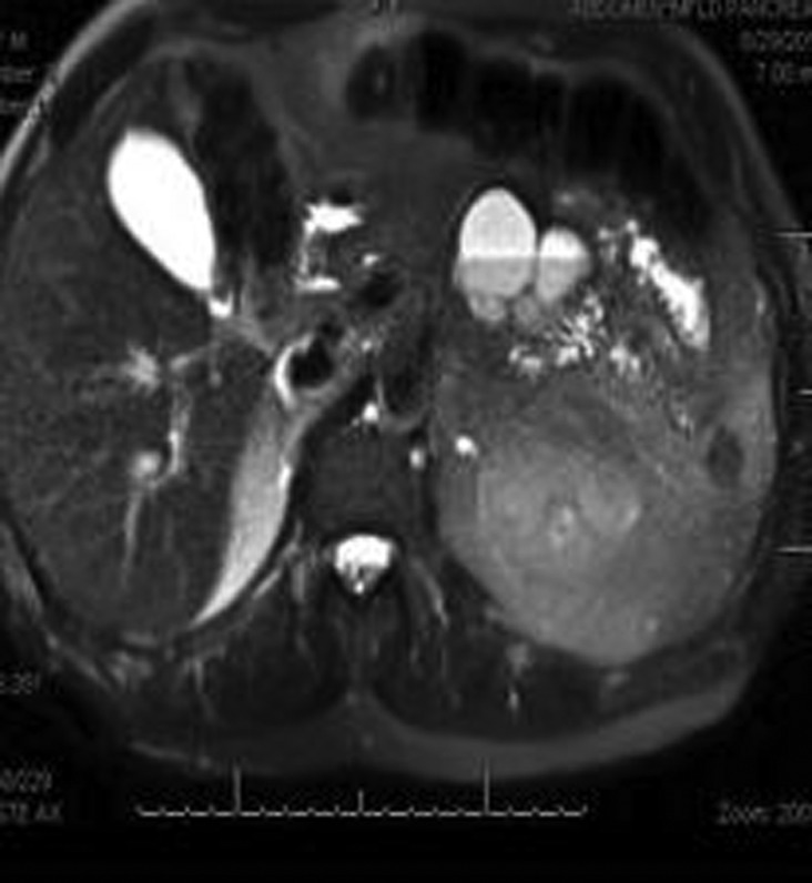
A 73-year-old man presented with a 5-month history of intermittent nausea, vomiting, central abdominal discomfort and a 17-pound weight loss over the past year. Laboratory testing, including a complete blood count with differential, liver function testing, amylase and lipase studies were normal. A CT scan showed a bilobed cystic lesion inferior to the body of the pancreas. An endoscopic ultrasound revealed a 5.3×3.9 cm, anechoic, bilobed cystic lesion, extrinsic to the body of the pancreas with a 1-2 mm septation and a normal pancreas. Fine-needle aspiration revealed a milky-white aspirate with negative cytology. Laboratory assessment of the cystic aspirant revealed carcinoembryonic antigen 1.7 ng/mL, amylase 148 units/L, cholesterol 300 mg/dL, and carbohydrate antigen 19-9 3 units/mL. He underwent resection of the mass, with the histopathology confirming a diagnosis of peripancreatic lymphangioma. He did well after the surgery with interval resolution of his symptoms.
Figures


Similar articles
-
Pancreas cystic lymphangioma diagnosed with EUS-FNA.JOP. 2012 May 10;13(3):282-4. JOP. 2012. PMID: 22572132
-
Pancreatic cystic lymphangioma in a 6-year-old girl, diagnosed by endoscopic ultrasound (EUS) fine needle aspiration.Endoscopy. 2011;43 Suppl 2 UCTN:E61-2. doi: 10.1055/s-0030-1256081. Epub 2011 Feb 1. Endoscopy. 2011. PMID: 21287455 No abstract available.
-
Recurrence of a pancreatic cystic lymphangioma after diagnosis and complete drainage by endoscopic ultrasound with fine-needle aspiration.JOP. 2013 May 10;14(3):280-2. doi: 10.6092/1590-8577/1347. JOP. 2013. PMID: 23669480
-
Role of endosocopic ultrasound in the diagnosis of cystic tumours of the pancreas.Dig Liver Dis. 2008 Nov;40(11):847-53. doi: 10.1016/j.dld.2008.03.019. Epub 2008 May 22. Dig Liver Dis. 2008. PMID: 18499542 Review.
-
Anaplastic carcinoma of the pancreas diagnosed by endoscopic ultrasound-guided fine-needle aspiration: a case report and review of the literature.J Med Case Rep. 2018 May 31;12(1):152. doi: 10.1186/s13256-018-1615-1. J Med Case Rep. 2018. PMID: 29848384 Free PMC article. Review.
Cited by
-
Rare Case of Pancreatic Cystic Lymphangioma.Intern Med. 2018 Mar 15;57(6):813-817. doi: 10.2169/internalmedicine.9445-17. Epub 2017 Nov 20. Intern Med. 2018. PMID: 29151529 Free PMC article.
References
-
- Paal E, Thompson LD, Heffess CS. A clinicopathological and a immunohistochemical study of ten pancreatic lymphangiomas and a review of the literature. Cancer 1998;2013:2150–8 - PubMed
-
- Wayne ER, Burrington JD, Bailey WC, et al. Retroperitoneal lymphangioma: an unusual case of acute surgical abdomen. J. Pediatric Surg 1973;2013:831–2 - PubMed
Publication types
MeSH terms
Substances
LinkOut - more resources
Full Text Sources
Other Literature Sources
Medical
Miscellaneous