. 2013 Dec;154(12):4627-39.
doi: 10.1210/en.2013-1483. Epub 2013 Oct 3.

The acid-secreting parietal cell as an endocrine source of Sonic Hedgehog during gastric repair

Affiliations

The acid-secreting parietal cell as an endocrine source of Sonic Hedgehog during gastric repair

Amy C Engevik et al. Endocrinology. 2013 Dec.

Abstract

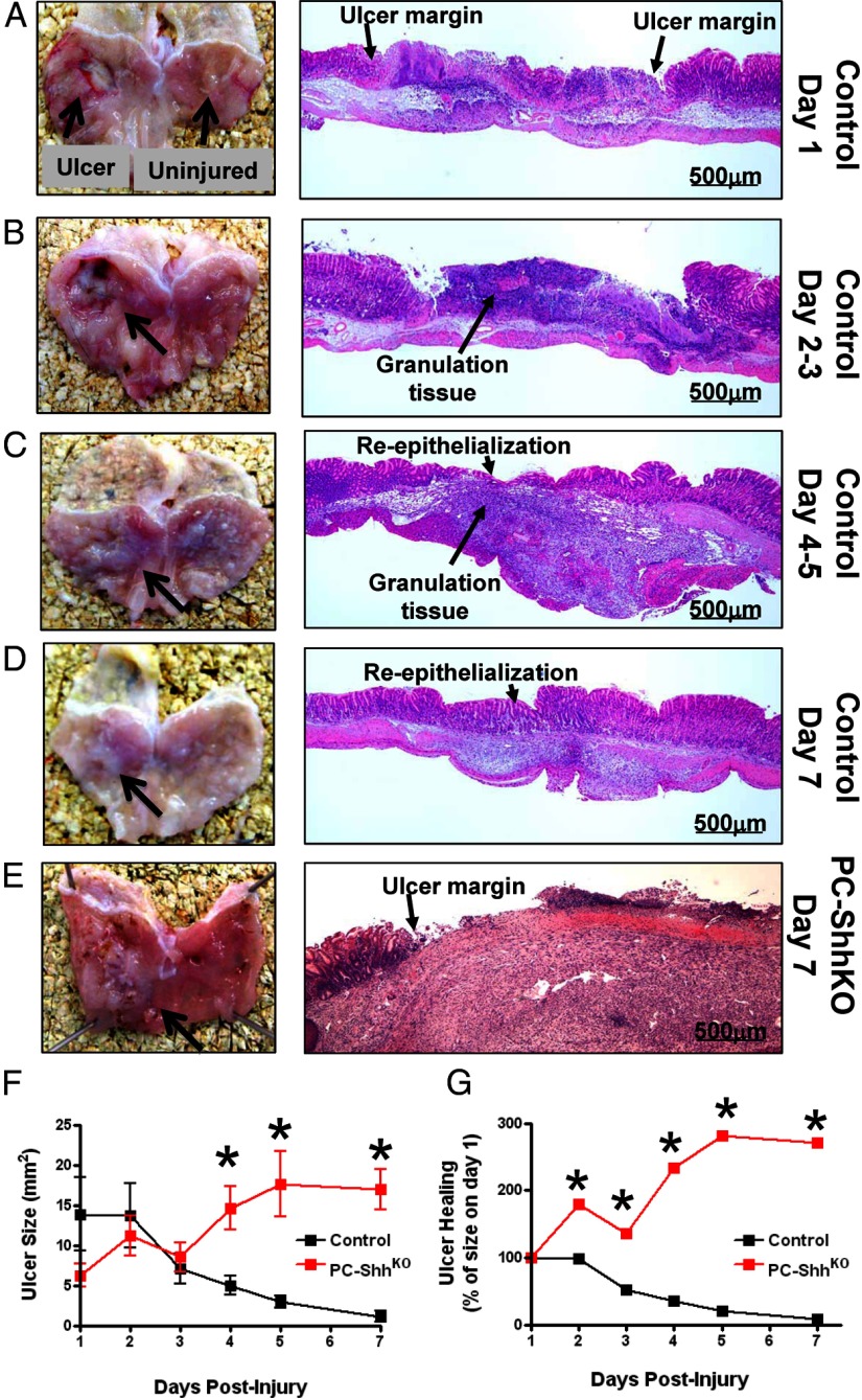
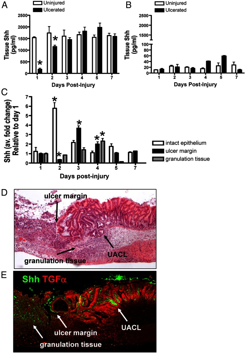
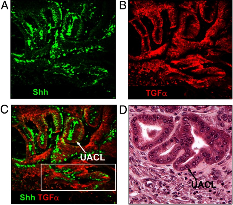
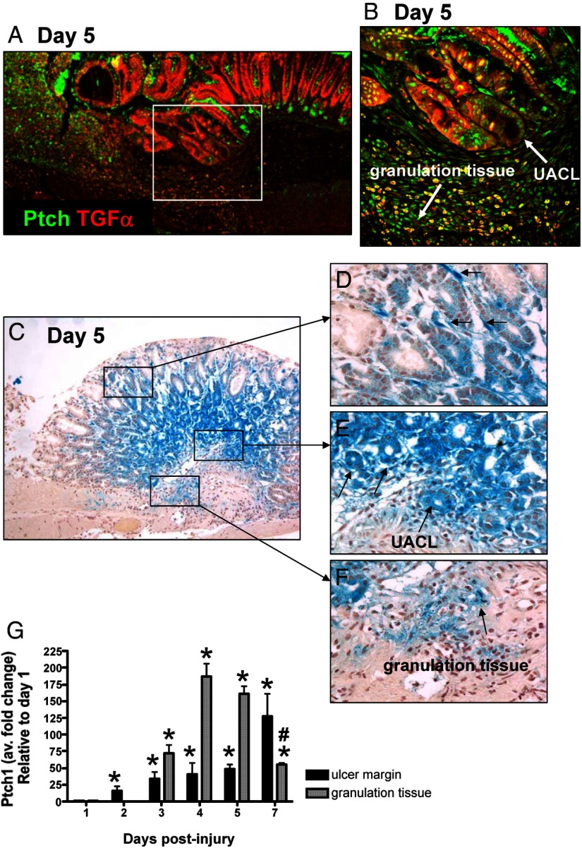
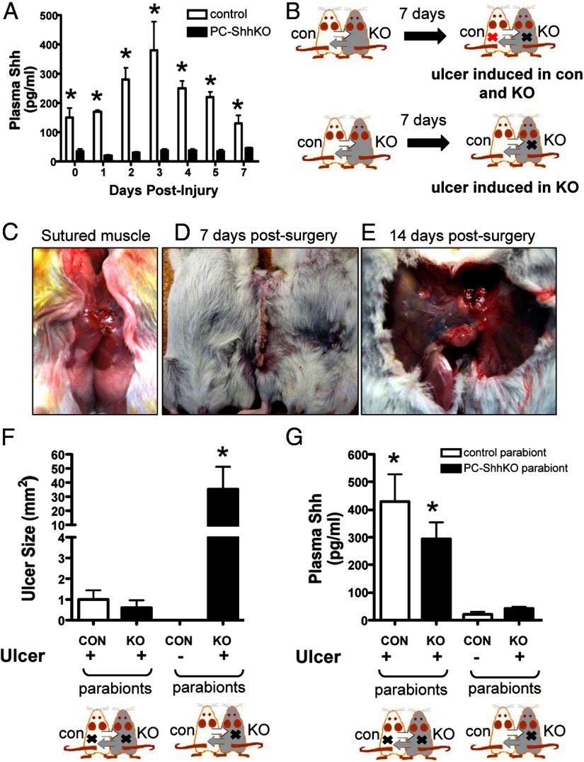
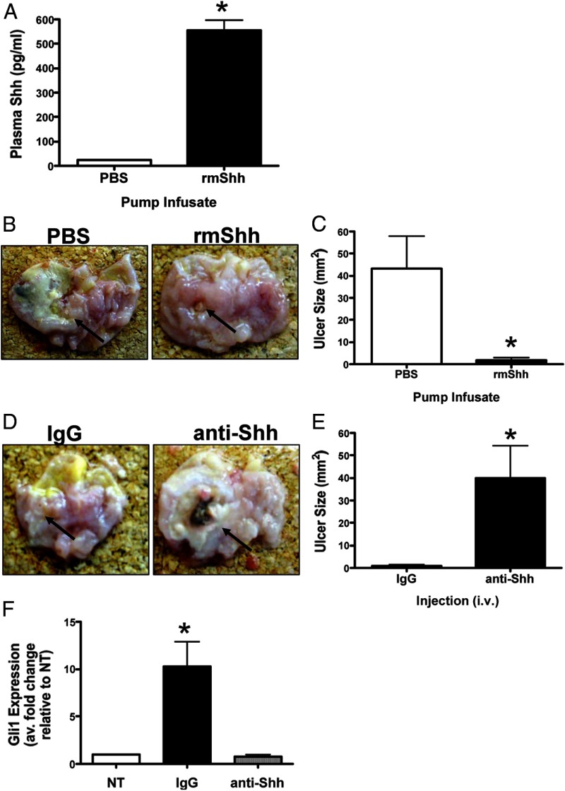
Sonic Hedgehog (Shh) has been shown to regulate wound healing in various tissues. Despite its known function in tissue regeneration, the role of Shh secreted from the gastric epithelium during tissue repair in the stomach remains unknown. Here we tested the hypothesis that Shh secreted from the acid-secreting parietal cell is a fundamental circulating factor that drives gastric repair. A mouse model expressing a parietal cell-specific deletion of Shh (PC-ShhKO) was generated using animals bearing loxP sites flanking exon 2 of the Shh gene (Shh(flx/flx)) and mice expressing a Cre transgene under the control of the H(+),K(+)-ATPase β-subunit promoter. Shh(flx/flx), the H(+),K(+)-ATPase β-subunit promoter, and C57BL/6 mice served as controls. Ulcers were induced via acetic acid injury. At 1, 2, 3, 4, 5, and 7 days after the ulcer induction, gastric tissue and blood samples were collected. Parabiosis experiments were used to establish the effect of circulating Shh on ulcer repair. Control mice exhibited an increased expression of Shh in the gastric tissue and plasma that correlated with the repair of injury within 7 days after surgery. PC-ShhKO mice showed a loss of ulcer repair and reduced Shh tissue and plasma concentrations. In a parabiosis experiment whereby a control mouse was paired with a PC-ShhKO littermate and both animals subjected to gastric injury, a significant increase in the circulating Shh was measured in both parabionts. Elevated circulating Shh concentrations correlated with the repair of gastric ulcers in the PC-ShhKO parabionts. Therefore, the acid-secreting parietal cell within the stomach acts as an endocrine source of Shh during repair.


Figures

Figure 1.
Figure 1.
Gross morphological and histological changes in control and PC-ShhKO mice in response to acetic acid-induced injury within the stomach. Gross morphology and H&E stains of control stomachs collected on days 1 (A), 2–3 (B), 4–5 (C), and 7 (D) postulcer induction depicting sites of ulcerated tissue and uninjured tissue used in experimental procedures. E, Gross morphology and H&E stain of PC-ShhKO stomach collected 7 days after the ulcer induction. F and G, Ulcer sizes for control and PC-ShhKO mice 1, 2, 3, 4, 5, and 7 days after the ulcer induction demonstrating disrupted ulcer repair in PC-ShhKO compared with complete healing in control mice (n = 5–17 mice per time point). *, P < .05 compared with control ulcer size.
Figure 2.
Figure 2.
Changes in Shh gastric tissue concentrations and gene expression. Changes in Shh tissue concentrations in gastric tissue collected from the stomachs of control (A) and PC-ShhKO (B) mice 1, 2, 3, 4, 5, and 7 days after acetic acid-induced injury. Data are expressed as the mean ± SEM with three to five mice per time point. *, P < .05 compared with uninjured gastric tissue. C, qRT-PCR was performed using RNA collected from intact epithelium, ulcer margin, and granulation tissue using LCM. Data are expressed as the mean ± SEM relative to day 1 with three to five mice per time point. *, P < .05 compared with day 1. D, H&E of control mouse stomach collected 5 days after acetic acid-induced injury showing a clear ulcer margin and infiltrating immune cells forming the granulation tissue. E, Immunofluorescence staining using an anti-Shh and anti-TGFα antibodies of a stomach section collected from a control mouse 5 days after acetic acid-induced injury. Shh expression (green) was observed at the ulcer margin and granulation tissue. Shh expression (green) also colocalized with TGFα-positive (red) UACL.
Figure 3.
Figure 3.
Expression of Shh within the UACL. Immunofluorescence staining using an anti-Shh and anti-TGFα antibodies of stomach sections collected from a control mouse 5 days after an acetic acid-induced injury. A, Shh expression (green) colocalized with TGFα-positive (red) UACL at the ulcer margin and within the granulation tissue (B). Merged image is shown in C. D, H&E demonstrating the presence of the ulcer-associated cell lineage at the base of regenerating glands at the ulcer margin and within the granulation tissue.
Figure 4.
Figure 4.
Expression of Ptch within the healing zone of the injured stomach. A, Immunofluorescence staining using an anti-Ptch and anti-TGFα antibodies of stomach sections collected from a control mouse 5 days after acetic acid-induced injury. Ptch expression (green) colocalized with TGFα-positive (red) UACL at the ulcer margin and within the granulation tissue. Higher magnification is shown in B. C, β-Galactosidase activity in the gastric mucosa of control mice 5 days after the acetic acid-induce injury using the heterozygote B61(29-Ptch1tm1Mps/J Ptch reporter mice). X-gal staining demonstrating Ptch1 expression shown within the mesenchyme (D), at the ulcer margin (E) and within the granulation tissue (F). G, qRT-PCR was performed using RNA collected from the ulcer margin and granulation tissue using LCM. Data are expressed as the mean ± SEM relative to day 1 with three to four mice per time point. *, P < .05 compared with day 1; #, P < .05 compared with days 4 and 5.
Figure 5.
Figure 5.
Changes in circulating Shh concentrations and ulcer sizes in control and PC-ShhKO parabionts. A, Changes in circulating Shh concentrations in blood collected from control and PC-ShhKO mice at 0, 1, 2, 3, 4, 5, and 7 days after acetic acid-induced injury. Data are expressed as the mean ± SEM with three to five mice per time point. *, P < .05 compared with control day 0. B, Design of the parabiosis experiments using control (CON) and PC-ShhKO (KO) parabionts. Surgery was performed to pair control and PC-ShhKO mice to establish a common circulation. An incision was made to expose the abdominal cavity wall and skin was pulled away from the muscle layer. C, A small incision was made below the diaphragm of each mouse, and the muscle layer of the two abdominal cavities were sutured together to form an opening between the abdominal cavities of each mouse. D, Wound clips were used to attach the skin between the two mice. E, Abdominal cavities were fused 14 days after parabiosis surgery. F, Ulcer size 7 days after the injury in control (CON) and PC-ShhKO (KO) parabionts in which both control and PC-ShhKO mice received an ulcer or parabionts in which only PC-ShhKO mice received ulcers. G, Shh concentration in plasma collected from control (CON) and PC-ShhKO (KO) parabionts. Data are expressed as the mean ± SEM with three to five pairs. *, P < .05 compared with parabionts with ulcers in both control and PC-ShhKO mice.
Figure 6.
Figure 6.
Changes in ulcer size in PC-ShhKO mice infused with exogenous Shh. A, Changes in circulating Shh concentrations in PC-ShhKO mice infused with either PBS or rmShh for 7 days after acetic acid-induced injury. B, Gross morphology of stomachs collected from PBS or rmShh infused PC-ShhKO mice. Arrows indicate ulcerated area. C, Changes in PC-ShhKO mouse ulcer size in response to either PBS or rmShh infusion for 7 days after acetic acid-induced injury. D, Gross morphology of stomachs collected from IgG or anti-Shh antibody-injected control mice. Arrows indicate ulcerated area. E, Changes in control mouse ulcer size in response to either IgG or anti-Shh antibody injections for 7 days after acetic acid-induced injury. Data are expressed as the mean ± SEM with five eight mice per group. *, P < .05 compared with PBS-infused or IgG-injected mice. F, Changes in Gli1 expression in Hedgehog-responsive cell line C3H10T1/2 in response to serum collected from either IgG- or anti-Shh antibody-injected control mice. *, P < .05 compared with no treatment (NT) (n = 5 serum samples collected from individual mice).
Figure 7.
Figure 7.
Expression of cytokine and growth factors in ulcerated stomachs of controls and PC-ShhKO mice. Expression of MIP-1α, MIP-2, IL-6, IL-1β, VEGF, and TGFβ using a Luminex multiplex-based assay in control (A–F) and PC-ShhKO (G–L) mouse stomachs collected 1, 2, 3, 4, 5, and 7 days after ulcer induction. n = 4–8 mice/time point.

References

    1. Grassi M, Petraccia L, Mennuni G, et al. Changes, functional disorders, and diseases in the gastrointestinal tract of elderly. Nutr Hosp. 2011;26:659–668 - PubMed
    1. Feldman M, Cryer B, McArthur KE, Huet BA, Lee E. Effects of aging and gastritis on gastric acid and pepsin secretion in humans: a prospective study. Gastroenterology. 1996;110:1043–1052 - PubMed
    1. Pilotto A, Franceschi M, Leandro G, Di Mario F, Valerio G. The effect of Helicobacter pylori infection on NSAID-related gastroduodenal damage in the elderly. Eur J Gastroenterol Hepatol. 1997;9:951–956 - PubMed
    1. Somerville K, Faulkner G, Langman M. Non-steroidal anti-inflammatory drugs and bleeding peptic ulcer. Lancet. 1986;1:462–464 - PubMed
    1. Everhart JE. Recent developments in the epidemiology of Helicobacter pylori. Gastroenterol Clin North Am. 2000;29:559–578 - PubMed

Publication types

MeSH terms