. 2007 Mar;2(2):205-215.
doi: 10.2217/17460794.2.2.205.

Filovirus replication and transcription

Affiliations

Filovirus replication and transcription

Elke Mühlberger. Future Virol. 2007 Mar.

Abstract

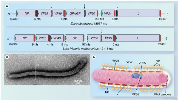
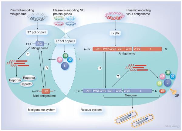
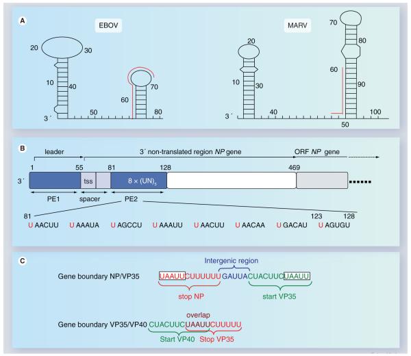
The highly pathogenic filoviruses, Marburg and Ebola virus, belong to the nonsegmented negative-sense RNA viruses of the order Mononegavirales. The mode of replication and transcription is similar for these viruses. On one hand, the negative-sense RNA genome serves as a template for replication, to generate progeny genomes, and, on the other hand, for transcription, to produce mRNAs. Despite the similarities in the replication/transcription strategy, filoviruses have evolved structural and functional properties that are unique among the nonsegmented negative-sense RNA viruses. Moreover, there are also striking differences in the replication and transcription mechanisms of Marburg and Ebola virus. This includes nucleocapsid formation, the structure of the genomic replication promoter, the protein requirement for transcription and the use of mRNA editing. In this article, the current knowledge of the replication and transcription strategy of Marburg and Ebola virus is reviewed, with focus on the observed differences.

Keywords: ebola virus; hemorrhagic fever; marburg virus; nonsegmented negative-sense RNA viruses; nucleocapsid complex; replication; reverse genetics; transcription.


Figures

Figure 1
Figure 1. Filovirus genome organization and structure
(A) Schematic diagram of Ebola virus (EBOV) and Marburg virus (MARV) genomes. The genes are depicted as boxes and nontranscribed regions as black bars (leader, trailer and intergenic regions). Transcription start signals are depicted as green triangles and stop signals as red bars. Gene overlaps are marked by arrows. The mRNA editing site within the EBOV GP gene is indicated by an asterisk. The length of the intergenic regions is shown below the scheme. Note that the EBOV VP24 gene contains two transcription stop signals. (B) Electron micrograph of an EBOV particle. (C) Schematic presentation of EBOV structure. The RNA genome is encapsidated by nucleocapsid proteins NP, VP35, VP30 and L. VP40 and VP24 are matrix proteins. GP trimers are inserted into the viral membrane. GP: Glycoprotein; L: RNA-dependent RNA polymerase; NP: Nucleoprotein; VP: Viral protein.
Figure 2
Figure 2. Hypothetical model of filovirus replication and transcription
Transcription results in the synthesis of seven monocistronic mRNAs that are capped and polyadenylated. The mRNAs are not encapsidated. For MARV transcription, NP, VP35 and L are sufficient. By contrast, EBOV transcription is dependent on the transcription activator VP30. Hypothetical model for EBOV transcription: (1) Transcription initiation: it is proposed that the polymerase consisting of L and VP35 initially binds to a single promoter site within the leader region and initiates transcription at the transcription start site of the first gene, the NP gene. Immediately after transcription initiation, the first 23 nucleotides of the nascent mRNA form a stable secondary structure (Figure 4A). In the absence of VP30, this RNA structure hampers movement of the polymerase along the RNA template and, consequently, the transcription complex pauses. (2) Transcription antitermination: the transcription block caused by the RNA secondary structure is abrogated by VP30 by a mechanism that is as yet unknown. It may be speculated that VP30 resolves the RNA structure or directs additional cofactors to the folded RNA. (3) Elongation and transcription reinitiation: transcription elongation and reinitiation at the following genes takes place independently of VP30, although each transcription start signal is involved in the formation of an RNA stem-loop structure [51]. Please note that the transcription start signal of MARV NP mRNA may also be involved in secondary structure formation. Nevertheless, MARV transcription occurs independently of VP30. mRNA editing has been observed only for EBOV GP mRNA [64,65]. Replication starts with the synthesis of a full-length positive-sense antigenome. The antigenome, in turn, serves as a template for the generation of progeny genomes. Both the genome and the antigenome are tightly encapsidated by the nucleocapsid proteins. NP, VP35 and L are sufficient to mediate MARV and EBOV replication. Since the antigenomic replication promoter is considered to be stronger than the genomic promoter, production of genomic RNA occurs more efficiently, as indicated by the blue arrow. Replication is performed by three proteins, NP, VP35 and L. Blue: negative-sense RNA; Red: positive-sense RNA. EBOV: Ebola virus; GP: Glycoprotein; L: RNA-dependent RNA polymerase; MARV: Marburg virus; NP: Nucleoprotein; VP: Viral protein.
Figure 3
Figure 3. Schematic diagram of filovirus reverse genetics systems
Minigenome system (left): cells are transfected with plasmids encoding the nucleocapsid proteins along with a plasmid encoding a virus-specific minigenome. The minigenome consists of a RG flanked by the 3′ and 5′ ends of the viral genome containing the signals for replication, encapsidation and transcription. The minigenome is T and R by the nucleocapsid proteins, leading to RG expression (reporter). Rescue system (right): here, cells are transfected with plasmids encoding the nucleocapsid proteins, along with a plasmid encoding a full-length copy of the positive-sense viral antigenome. The antigenome is first R by the nucleocapsid proteins resulting in the generation of a negative-sense genome. The genome serves as a template for replication and transcription, leading to viral mRNA production and subsequent protein synthesis. Finally, the genomes are packaged by the viral proteins and released into mature infectious virus particles. The nucleocapsid protein genes, minigenomes and full-length antigenomes can be cloned under the control of different promoters (T7 pol, pol I and pol II). Negative-sense RNA is indicated in blue, positive-sense RNA in red. GP: Glycoprotein; L: RNA-dependent RNA polymerase; NP: Nucleoprotein; pol I: RNA polymerase I; pol II: RNA polymerase II; R: Replicated; RG: Reporter gene; T7 pol: T7 RNA polymerase; T: Transcribed; VP: Viral protein.
Figure 4
Figure 4. Cis-acting elements on filovirus genomes
(A) RNA secondary structure formation at the 3′ terminus of MARV and EBOV genomes. The first predicted RNA secondary structure is located within the leader region of each genome, the second RNA stem-loop structure is formed by the respective first transcription start site and downstream-located sequences. The red lines running along the side of the stem-loops identify the transcription start signals. Predicted panhandle structures formed by complementary regions of the 3′ and 5′ ends are not shown. Nucleotide numbers refer to the genomic RNA of Zaire ebolavirus, strain Mayinga (accession number AF086833) and Lake Victoria marburgvirus, strain Musoke (accession number DQ217792), respectively. (B) Structure of the Zaire ebolavirus genomic replication promoter. The promoter is located at the 3′ end of the genome. It is bipartite consisting of PE 1 and 2. PE 1 and 2 are depicted as blue boxes. PE 1 and 2 are separated by a spacer (light blue) that contains the transcription start signal of the NP gene. PE 1 spans the leader region, PE 2 is located within the nontranslated region of the NP gene and consists of a stretch of eight UN5 hexamers. The ORF of the NP gene is depicted in light gray. The nucleotide sequence of PE 2 is shown below the scheme. Conserved hexameric U residues are indicated in red. (C) Two typical gene boundaries within the EBOV genome. Transcription start signals are shown in green, transcription stop signals in red. Intergenic region is shown in blue, gene overlap in purple. A conserved pentamer, which is part of the transcription start signals as well as of the transcription stop signals, is boxed. EBOV: Ebola virus; MARV: Marburgvirus; NP: Nucleoprotein; ORF: Open-reading frame; PE: Promoter element; tss: Transcription start signal; VP: Viral protein.

Similar articles

Cited by

References

    1. Hensley LE, Jones SM, Feldmann H, Jahrling PB, Geisbert TW. Ebola and Marburg viruses: pathogenesis and development of countermeasures. Curr. Mol. Med. 2005;5:761–772. - PubMed
    1. Feldmann H, Geisbert TW, Jahrling PB, et al. In: Virus Taxonomy, Eighth Report of the International Committee on Taxonomy of Viruses. Fauquet CM, Mayo MA, Maniloff J, Desselberger U, Ball LA, editors. Elsevier/Academic Press; San Diego, USA: 2005. pp. 645–653.
    1. Wang L, Harcourt BH, Yu M, et al. Molecular biology of Hendra and Nipah viruses. Microbes Infect. 2001;3:279–287. - PubMed
    1. Towner JS, Khristova ML, Sealy TK, et al. Marburgvirus genomics and association with a large hemorrhagic fever outbreak in Angola. J. Virol. 2006;80:6497–6516. - PMC - PubMed
    1. Sanchez A, Rollin PE. Complete genome sequence of an Ebola virus (Sudan species) responsible for a 2000 outbreak of human disease in Uganda. Virus Res. 2005;113:16–25. - PubMed

LinkOut - more resources