Review
. 2013 Oct;4(5):447-64.
doi: 10.1515/bmc-2013-0023.

NDE1 and NDEL1: twin neurodevelopmental proteins with similar 'nature' but different 'nurture'

Affiliations
Review

NDE1 and NDEL1: twin neurodevelopmental proteins with similar 'nature' but different 'nurture'

Nicholas J Bradshaw et al. Biomol Concepts. 2013 Oct.

Abstract

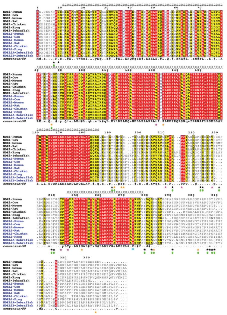
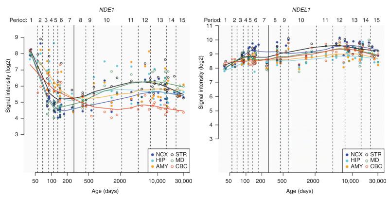
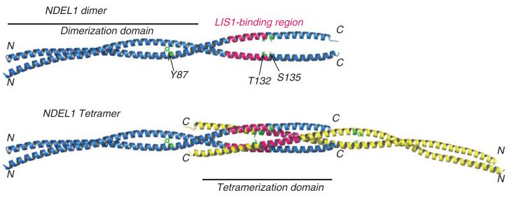
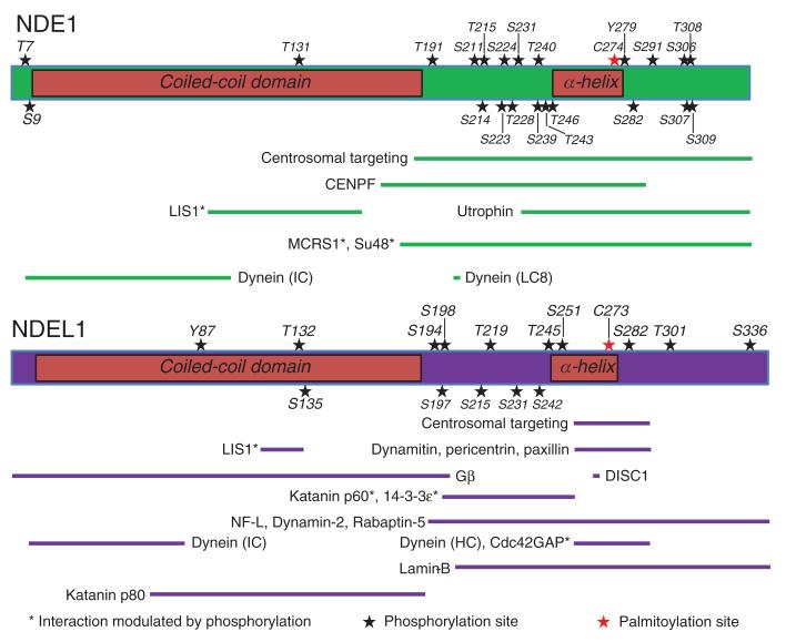
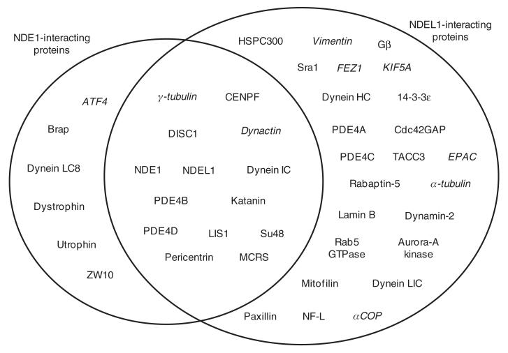
Nuclear distribution element 1 (NDE1, also known as NudE) and NDE-like 1 (NDEL1, also known as Nudel) are paralogous proteins essential for mitosis and neurodevelopment that have been implicated in psychiatric and neurodevelopmental disorders. The two proteins possess high sequence similarity and have been shown to physically interact with one another. Numerous lines of experimental evidence in vivo and in cell culture have demonstrated that these proteins share common functions, although instances of differing functions between the two have recently emerged. We review the key aspects of NDE1 and NDEL1 in terms of recent advances in structure elucidation and cellular function, with an emphasis on their differing mechanisms of post-translational modification. Based on a review of the literature and bioinformatics assessment, we advance the concept that the twin proteins NDE1 and NDEL1, while sharing a similar 'nature' in terms of their structure and basic functions, appear to be different in their 'nurture', the manner in which they are regulated both in terms of expression and of post-translational modification within the cell. These differences are likely to be of significant importance in understanding the specific roles of NDE1 and NDEL1 in neurodevelopment and disease.


Figures

Figure 1
Figure 1
Multiple sequence alignment of NDE1 and NDEL1 orthologs. Orthologs of NDE1 and NDEL1 across vertebrate species were identified using the UCSC Genome Browser (http://genome-euro.ucsc.edu). These were aligned initially using Clustal Omega (108, 109) with further manual editing for optimum alignment. The sequence numbering at the top of each alignment block corresponds to the sequence of human NDE1. Conserved residues are colored with a red background; conservatively substituted residues are shown with a yellow background. The location of the N-terminal coiled-coil α-helical domain (residue ~10–185) and the predicted C-terminal α-helix (residue ~247–278) are shown above the alignment. Consensus amino acids are shown at the bottom of the each alignment block (uppercase is strictly conserved; lowercase is consensus level > 0.5;! is I or V; $ is L or M; % is F or Y; = is N, D, Q or E). Despite 60% sequence identity there are at least 18 specific phosphorylation sites for each human protein – i.e., phosphorylation possible only in one protein. Some of these phosphorylation sites have been derived from high-throughput proteomic screening using mass spectrometry (90), while others have been experimentally verified in the cell. See the main text and Table 2 for more information. Those sites known to be specific to either NDE1 or NDEL1 are highlighted with a green filled oval under the corresponding star; black star=phosphorylation site in NDE1; orange star=phosphorylation site in NDEL1; pink star=phosphorylation site in both NDE1 and NDEL1; cyan block=palmitoylation site in both proteins. The figure was generated with ESPript v2.2 (111).
Figure 2
Figure 2
Differential expression of NDE1 and NDEL1 transcripts in the human brain across the lifetime. Change in NDE1 mRNA expression with time is shown in the left panel and NDEL1 in the right panel; developmental age shown on the x-axis in days; mRNA expression signal intensity (Log2) shown on the y-axis. While, expression of NDEL1 across the brain remains relatively steady, NDE1 shows a far more specialized pattern of expression. Abbreviations for brain regions used in figure: NCX, neocortex; STR, striatum; HIP, hippocampus; MD, mediodorsal nucleus of the thalamus; AMY, amygdala; CBC, cerebellar cortex. Data and figure accessed from the Human Brain Transcriptome project (http://hbatlas.org/pages/hbtd; used with permission) (79).
Figure 3
Figure 3
Coiled-coil crystal structure of NDEL1 with known post-translational modifications mapped. Only three known PTMs map onto the crystal structure of the NDEL1 coiled-coil domain fragment (residues 8–167; PDB ID: 2V71); these side-chains are shown in green and labeled on cartoon representations of the NDEL1 crystal structure, parallel dimer (top panel) and anti-parallel tetramer (bottom panel) (81); N- and C-termini for each chain and regions that facilitate dimerization and tetramerization are indicated. The essential LIS1-binding region on NDEL1 114–133 (96) is also shown (pink). Phosphorylation of NDE1 at T131 (directly equivalent to T132 of NDEL1) reduces its ability to interact with LIS1 (86). It can therefore be hypothesized that phosphorylation of T132 and/or S135 of NDEL1 may have similar effects on LIS1-binding. The Y87 site is highly solvent-exposed on the dimerization domain of coiled-coil structure and is thus readily accessible to kinases.
Figure 4
Figure 4
Domain architecture, post-translation modification and binding/functional regions of NDE1 and NDEL1. Schematics of NDE1 (top panel) and NDEL1 (bottom panel) drawn to scale depicting the N-terminal coiled-coil domain and the predicted C-terminal α-helix (labeled); the central flexible linker region connects these two structured regions, while another unstructured region lies at the extreme C-terminus of the proteins. Numerous PTMs (stars) reside within delineated protein-protein interaction sites, sub-cellular localization regions, or oligomerization domains with the vast majority located within predicted unstructured regions of the proteins. Minimal essential protein-binding sites from the literature are shown drawn to scale (NDE1: green bars; NDEL1: purple bars). References for each NDE1 interaction site are as follows: Centrosomal targeting (98); Centromere protein F (CENPF)/Mitosin/LEK1 (112); LIS1 (4); Utrophin (66); MCRS1/p78 (99); Su48/DBZ/ZNF365 (98); Dynein IC (89); Dynein (LC8) (49). References for each NDEL1 interaction site are as follows: Centrosomal targeting (113); LIS1 (96); Dynamitin (50); Pericentrin/Kendrin (113); Paxillin (114); G-protein β (Gβ) (115); DISC1 (28); Katanin p60 and p80 (68); 14-3-3ε (100); Neurofilament (NF)-L (72); Dynamin-2 (75); Rabaptin-5 (78); Dynein intermediate chain (IC) (89); Dynein heavy chain (HC) (1); Cdc42GAP (74); Lamin-B (63). Note: the palmitoylation site C273 (red star) on NDEL1 is also the site of endooligopeptidase activity (77). In the majority of cases, these sites have only been investigated for one of NDE1 or NDEL1; it is likely that some of them will be conserved between the two proteins. The effect of PTMs should be tested on protein conformation, stability, oligomerization, modulation of protein-protein interaction and cellular trafficking/targeting.
Figure 5
Figure 5
Protein interaction partners of NDE1 and NDEL1. Proteins in the left circle interact with NDE1, those in the right circle with NDEL1. Proteins in the intersecting region are known to interact with both proteins. In the majority of instances where a protein is listed as interacting with only one of NDE1/NDEL1, the reciprocal experiment to test for interaction with the other of NDEL1/NDE1 has not been published to the best of our knowledge. Proteins listed in italics are known to exist in a complex with NDE1/NDEL1 but do not, or have not yet been shown to, directly bind to it. Data are taken from the following papers (1-5, 27-29, 31-33, 48, 50, 53, 57, 58, 63, 65, 66, 68, 72, 74-76, 78, 80, 88, 90, 96-98, 100, 101, 113-121). In some cases the essential binding regions for these interaction partners have been delineated on NDE1 or NDEL1; these are shown schematically in Figure 4.

Similar articles

Cited by

References

    1. Sasaki S, Shionoya A, Ishida M, Gambello MJ, Yingling J, Wynshaw-Boris A, Hirotsune S. A LIS1/NUDEL/cytoplasmic dynein heavy chain complex in the developing and adult nervous system. Neuron. 2000;28:681–96. - PubMed
    1. Niethammer M, Smith DS, Ayala R, Peng J, Ko J, Lee MS, Morabito M, Tsai LH. NUDEL is a novel Cdk5 substrate that associates with LIS1 and cytoplasmic dynein. Neuron. 2000;28:697–711. - PubMed
    1. Kitagawa M, Umezu M, Aoki J, Koizumi H, Arai H, Inoue K. Direct association of LIS1, the lissencephaly gene product, with a mammalian homologue of a fungal nuclear distribution protein, rNUDE. FEBS Lett. 2000;479:57–62. - PubMed
    1. Feng Y, Olson EC, Stukenberg PT, Flanagan LA, Kirschner MW, Walsh CA. LIS1 regulates CNS lamination by interacting with mNudE, a central component of the centrosome. Neuron. 2000;28:665–79. - PubMed
    1. Sweeney KJ, Prokscha A, Eichele G. NudE-L, a novel Lis1-interacting protein, belongs to a family of vertebrate coiled-coil proteins. Mech Dev. 2001;101:21–33. - PubMed

Publication types

MeSH terms

LinkOut - more resources